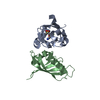
 Movie
Movie Controller


+ Open data

- Basic information
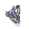
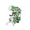
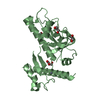
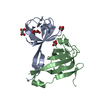
| Entry | Database: PDB / ID: 6fms | ||||||||||||
|---|---|---|---|---|---|---|---|---|---|---|---|---|---|
| Title | IMISX-EP of Se-LspA | ||||||||||||
 Components Components |
| ||||||||||||
 Keywords Keywords |  HYDROLASE / Serial crystallography / experimental phasing / in meso crystallization / in situ diffraction data collection / membrane protein structure / MEMBRANE PROTEIN HYDROLASE / Serial crystallography / experimental phasing / in meso crystallization / in situ diffraction data collection / membrane protein structure / MEMBRANE PROTEIN | ||||||||||||
| Function / homology |  Function and homology informationsignal peptidase II / signal peptide processing / aspartic-type endopeptidase activity / plasma membrane Function and homology informationsignal peptidase II / signal peptide processing / aspartic-type endopeptidase activity / plasma membraneSimilarity search - Function | ||||||||||||
| Biological species |   Pseudomonas aeruginosa (bacteria) Pseudomonas aeruginosa (bacteria)synthetic construct (others) | ||||||||||||
| Method | X-RAY DIFFRACTION / SYNCHROTRON / SAD / Resolution: 3 Å | ||||||||||||
 Authors Authors | Huang, C.-Y. / Olieric, V. / Howe, N. / Warshamanage, R. / Weinert, T. / Panepucci, E. / Vogeley, L. / Basu, S. / Diederichs, K. / Caffrey, M. / Wang, M. | ||||||||||||
| Funding support |  Ireland, Ireland,  Switzerland, 2items Switzerland, 2items
| ||||||||||||
 Citation Citation | Journal: Commun Biol / Year: 2018 Title: In situ serial crystallography for rapid de novo membrane protein structure determination. Authors: Huang, C.Y. / Olieric, V. / Howe, N. / Warshamanage, R. / Weinert, T. / Panepucci, E. / Vogeley, L. / Basu, S. / Diederichs, K. / Caffrey, M. / Wang, M. | ||||||||||||
| History |
|
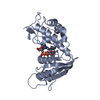
- Structure visualization
| Structure viewer | Molecule: MolmilJmol/JSmol |
|---|
- Downloads & links
-Download
| PDBx/mmCIF format | 6fms.cif.gz | 155 KB | Display | PDBx/mmCIF format |
|---|---|---|---|---|
| PDB format | pdb6fms.ent.gz | 121.9 KB | Display | PDB format |
| PDBx/mmJSON format | 6fms.json.gz | Tree view | PDBx/mmJSON format | |
| Others |  Other downloads Other downloads |
-Validation report
| Arichive directory | https://data.pdbj.org/pub/pdb/validation_reports/fm/6fmsftp://data.pdbj.org/pub/pdb/validation_reports/fm/6fms | HTTPS FTP |
|---|
-Related structure data
| Related structure data | 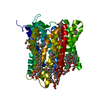 6fmrC  6fmtC  6fmvC  6fmwC  6fmxC 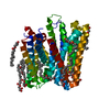 6fmyC C: citing same article ( |
|---|---|
| Similar structure data |


-Links
 PDBj
PDBj


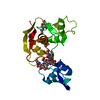
- Assembly
| Deposited unit | 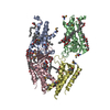
| ||||||||
|---|---|---|---|---|---|---|---|---|---|
| 1 | 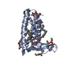
| ||||||||
| 2 | 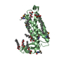
| ||||||||
| 3 | 
| ||||||||
| 4 | 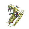
| ||||||||
| Unit cell |
|
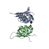
-Components
| #1: Protein | Mass: 21287.877 Da / Num. of mol.: 4 Source method: isolated from a genetically manipulated source Source: (gene. exp.) Pseudomonas aeruginosa (strain ATCC 15692 / DSM 22644 / CIP 104116 / JCM 14847 / LMG 12228 / 1C / PRS 101 / PAO1) (bacteria)Gene: lspA, ls, PA4559 / Production host: Escherichia coli BL21(DE3) (bacteria) / References: UniProt: Q9HVM5, signal peptidase II#2: Protein/peptide |   Mass: 673.838 Da / Num. of mol.: 4 / Source method: obtained synthetically / Source: (synth.) synthetic construct (others) / References: Globomycin Mass: 673.838 Da / Num. of mol.: 4 / Source method: obtained synthetically / Source: (synth.) synthetic construct (others) / References: Globomycin#3: Chemical | ChemComp-OLC / (   Mass: 356.540 Da / Num. of mol.: 22 / Source method: obtained synthetically / Formula: C21H40O4 Mass: 356.540 Da / Num. of mol.: 22 / Source method: obtained synthetically / Formula: C21H40O4#4: Water | ChemComp-HOH / | Water Mass: 18.015 Da / Num. of mol.: 10 / Source method: isolated from a natural source / Formula: H2O Mass: 18.015 Da / Num. of mol.: 10 / Source method: isolated from a natural source / Formula: H2O |
|---|
-Experimental details
-Experiment
| Experiment | Method: X-RAY DIFFRACTION / Number of used crystals: 1 |
|---|
- Sample preparation
| Crystal | Density Matthews: 3.78 Å3/Da / Density % sol: 67.49 % |
|---|---|
| Crystal grow | Temperature: 293 K / Method: lipidic cubic phase Details: 35 % PEG 400, 100 mM MES, pH 5.6, and 100 mM ammonium phosphate monobasic |
-Data collection
| Diffraction | Mean temperature: 100 K |
|---|---|
| Diffraction source | Source: SYNCHROTRON / Site: SLS / Beamline: X06SA / Wavelength: 0.97858 Å |
| Detector | Type: DECTRIS EIGER X 16M / Detector: PIXEL / Date: Feb 16, 2015 |
| Radiation | Protocol: SINGLE WAVELENGTH / Monochromatic (M) / Laue (L): M / Scattering type: x-ray |
| Radiation wavelength | Wavelength: 0.97858 Å / Relative weight: 1 |
| Reflection | Resolution: 3→46.27 Å / Num. obs: 41032 / % possible obs: 99.9 % / Redundancy: 22.7 % / CC1/2: 0.99 / Rrim(I) all: 0.31 / Net I/σ(I): 9.14 |
| Reflection shell | Resolution: 3→3.08 Å / Redundancy: 22.7 % / Mean I/σ(I) obs: 1.45 / Num. unique obs: 3031 / CC1/2: 0.4 / Rrim(I) all: 3.46 / % possible all: 99.8 |
- Processing
| Software |
| ||||||||||||||||
|---|---|---|---|---|---|---|---|---|---|---|---|---|---|---|---|---|---|
| Refinement | Method to determine structure: SAD / Resolution: 3→46.268 Å / Cross valid method: THROUGHOUT
| ||||||||||||||||
| Refinement step | Cycle: LAST / Resolution: 3→46.268 Å
|
