Component-ID: 0 / Beg auth comp-ID: VAL / Beg label comp-ID: VAL / End auth comp-ID: HIS / End label comp-ID: HIS / Refine code: 0 / Auth seq-ID: 38 - 430 / Label seq-ID: 38 - 430
Dom-ID
Ens-ID
Auth asym-ID
Label asym-ID
1
1
A
A
2
1
B
B
1
2
A
A
2
2
C
C
1
3
A
A
2
3
D
D
1
4
A
A
2
4
E
E
1
5
A
A
2
5
F
F
1
6
B
B
2
6
C
C
1
7
B
B
2
7
D
D
1
8
B
B
2
8
E
E
1
9
B
B
2
9
F
F
1
10
C
C
2
10
D
D
1
11
C
C
2
11
E
E
1
12
C
C
2
12
F
F
1
13
D
D
2
13
E
E
1
14
D
D
2
14
F
F
1
15
E
E
2
15
F
F
NCS ensembles :
ID
1
2
3
4
5
6
7
8
9
10
11
12
13
14
15
-
Components
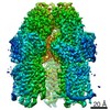
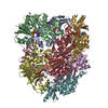
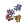
#1: Protein
Glycosidehydrolase, GH17family /
Mass: 48953.215 Da / Num. of mol.: 6 Source method: isolated from a genetically manipulated source Source: (gene. exp.) Formosa sp. Hel1_33_131 (bacteria) / Gene: FORMB_24720 / Production host: Escherichia coli BL21(DE3) (bacteria) / References: UniProt: A0A1D7Y259
Resolution: 2.6→104.32 Å / Cor.coef. Fo:Fc: 0.923 / Cor.coef. Fo:Fc free: 0.892 / SU B: 11.151 / SU ML: 0.225 / Cross valid method: THROUGHOUT / ESU R: 1.151 / ESU R Free: 0.308 / Details: HYDROGENS HAVE BEEN ADDED IN THE RIDING POSITIONS
Rfactor
Num. reflection
% reflection
Selection details
Rfree
0.23881
4247
4.9 %
RANDOM
Rwork
0.20461
-
-
-
obs
0.20629
83120
99.7 %
-
Solvent computation
Ion probe radii: 0.8 Å / Shrinkage radii: 0.8 Å / VDW probe radii: 1.2 Å
 Movie
Movie Controller

 Yorodumi
 Open data
 Basic information
 Components
 Keywords
 Function and homology information
 Formosa sp. Hel1_33_131 (bacteria)
 Authors
 Germany, 1items
  Citation
 Structure visualization
 Downloads & links
 Other downloads


 PDBj
PDBj Assembly
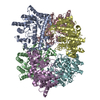

 Mass: 40.078 Da / Num. of mol.: 2 / Source method: obtained synthetically / Formula: Ca
 Mass: 18.015 Da / Num. of mol.: 863 / Source method: isolated from a natural source / Formula: H2O
 Sample preparation
 Processing