Movie
Movie Controller

[English] 日本語
 Yorodumi
- PDB-6e9d: Sub-2 Angstrom Ewald Curvature-Corrected Single-Particle Cryo-EM ... -
+ Open data

- Basic information
| Entry | Database: PDB / ID: 6e9d | ||||||||||||
|---|---|---|---|---|---|---|---|---|---|---|---|---|---|
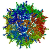
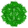
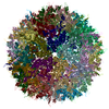
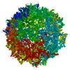
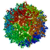
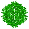
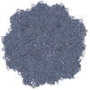
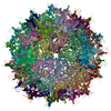
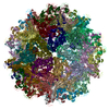
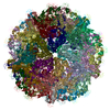
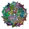
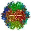
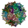
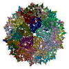
| Title | Sub-2 Angstrom Ewald Curvature-Corrected Single-Particle Cryo-EM Reconstruction of AAV-2 L336C | ||||||||||||
 Components Components | Capsid protein VP1 | ||||||||||||
 Keywords Keywords | VIRUS LIKE PARTICLE / AAV-2 L336C / Gene therapy / Ewald sphere curvature correction / Per particle CTF | ||||||||||||
| Function / homology |  Function and homology information Function and homology informationpermeabilization of host organelle membrane involved in viral entry into host cell / symbiont entry into host cell via permeabilization of inner membrane / host cell nucleolus / T=1 icosahedral viral capsid / clathrin-dependent endocytosis of virus by host cell / structural molecule activity / virion attachment to host cell Similarity search - Function | ||||||||||||
| Biological species |  Adeno-associated virus 2 Srivastava/1982 Adeno-associated virus 2 Srivastava/1982 | ||||||||||||
| Method | ELECTRON MICROSCOPY / single particle reconstruction / cryo EM / Resolution: 1.86 Å | ||||||||||||
 Authors Authors | Tan, Y.Z. / Aiyer, S. / Mietzsch, M. / Hull, J.A. / McKenna, R. / Baker, T.S. / Agbandje-McKenna, M. / Lyumkis, D. | ||||||||||||
| Funding support |  United States, 3items United States, 3items
| ||||||||||||
 Citation Citation | Journal: Nat Commun / Year: 2018 Title: Sub-2 Å Ewald curvature corrected structure of an AAV2 capsid variant. Authors: Yong Zi Tan / Sriram Aiyer / Mario Mietzsch / Joshua A Hull / Robert McKenna / Joshua Grieger / R Jude Samulski / Timothy S Baker / Mavis Agbandje-McKenna / Dmitry Lyumkis / Abstract: Single-particle cryogenic electron microscopy (cryo-EM) provides a powerful methodology for structural biologists, but the resolutions typically attained with experimentally determined structures ...Single-particle cryogenic electron microscopy (cryo-EM) provides a powerful methodology for structural biologists, but the resolutions typically attained with experimentally determined structures have lagged behind microscope capabilities. Here, we exploit several technical advances to improve resolution, including per-particle contrast transfer function (CTF) refinement and correction for Ewald sphere curvature. The latter is demonstrated with several experimental samples and should become more standard as resolutions increase or at lower microscope accelerating voltages. The combined application of the described methods to micrographs recorded on a Titan Krios enables structure determination at ~1.86-Å resolution of an adeno-associated virus serotype 2 variant (AAV2), an important gene-delivery vehicle. The resulting structural details provide an improved model for understanding the biology of AAV that will guide future vector development for gene therapy. | ||||||||||||
| History |
|
- Structure visualization
| Movie |
Movie viewer |
|---|---|
| Structure viewer | Molecule: MolmilJmol/JSmol |
- Downloads & links
-Download
| PDBx/mmCIF format | 6e9d.cif.gz | 10.8 MB | Display | PDBx/mmCIF format |
|---|---|---|---|---|
| PDB format | pdb6e9d.ent.gz | Display | PDB format | |
| PDBx/mmJSON format | 6e9d.json.gz | Tree view | PDBx/mmJSON format | |
| Others |  Other downloads Other downloads |
-Validation report
| Arichive directory | https://data.pdbj.org/pub/pdb/validation_reports/e9/6e9dftp://data.pdbj.org/pub/pdb/validation_reports/e9/6e9d | HTTPS FTP |
|---|
-Related structure data
| Related structure data |  9012MC M: map data used to model this data C: citing same article ( |
|---|---|
| Similar structure data | |
| EM raw data | EMPIAR-10202 (Title: Sub-2 Å Single-Particle Cryo-EM Reconstruction of AAV2-L336C Data size: 5.7 TB Data #1: Unaligned, compressed, raw movie frames of AAV2-L336C [micrographs - multiframe] Data #2: MotionCor2 aligned, gain corrected, magnification anisotropy corrected, Fourier bin by 2 movie frames of AAV2-L336C [micrographs - multiframe] Data #3: MotionCor2 aligned, gain corrected, magnification anisotropy corrected, Fourier bin by 2, dose weighted, summed micrographs of AAV2-L336C [micrographs - single frame] Data #4: MotionCor2 aligned, gain corrected, magnification anisotropy corrected, Fourier bin by 2, not dose weighted, summed micrographs of AAV2-L336C [micrographs - single frame] Data #5: MotionCor2 aligned, gain corrected, magnification anisotropy corrected, Fourier bin by 2, not dose weighted, frames 5 to 19 summed micrographs of AAV2-L336C [micrographs - single frame] Data #6: Final particle stack of AAV2-L336C using frames 5 to 19 [micrographs - single frame] Data #7: Final particle stack of AAV2-L336C grouped into frames 5-9, 10-14 and 15-19, after rotational alignment [micrographs - single frame]) |


-Links
 PDBj
PDBj


- Assembly
| Deposited unit | 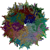
|
|---|---|
| 1 |
|
-Components
| #1: Protein | Mass: 82021.336 Da / Num. of mol.: 60 / Mutation: L336C Source method: isolated from a genetically manipulated source Source: (gene. exp.) Adeno-associated virus 2 Srivastava/1982Gene: VP1 / Cell line (production host): Sf9 / Production host:   Spodoptera frugiperda (fall armyworm) / References: UniProt: P03135 Spodoptera frugiperda (fall armyworm) / References: UniProt: P03135#2: Water | ChemComp-HOH / | Water Mass: 18.015 Da / Num. of mol.: 11820 / Source method: isolated from a natural source / Formula: H2O Mass: 18.015 Da / Num. of mol.: 11820 / Source method: isolated from a natural source / Formula: H2O |
|---|
-Experimental details
-Experiment
| Experiment | Method: ELECTRON MICROSCOPY |
|---|---|
| EM experiment | Aggregation state: PARTICLE / 3D reconstruction method: single particle reconstruction |
- Sample preparation
| Component | Name: Adeno-associated virus - 2 / Type: VIRUS / Entity ID: #1 / Source: RECOMBINANT | ||||||||||||||||||||
|---|---|---|---|---|---|---|---|---|---|---|---|---|---|---|---|---|---|---|---|---|---|
| Molecular weight | Value: 3.9 MDa / Experimental value: NO | ||||||||||||||||||||
| Source (natural) | Organism: Adeno-associated virus - 2 | ||||||||||||||||||||
| Source (recombinant) | Organism: Spodoptera frugiperda (fall armyworm) / Strain: Sf9 / Plasmid: pFastBac1-HM | ||||||||||||||||||||
| Details of virus | Empty: YES / Enveloped: NO / Isolate: STRAIN / Type: VIRUS-LIKE PARTICLE | ||||||||||||||||||||
| Natural host | Organism: Homo sapiens | ||||||||||||||||||||
| Virus shell | Name: Icosahedral capsidCapsid / Diameter: 250 nm / Triangulation number (T number): 1 | ||||||||||||||||||||
| Buffer solution | pH: 7.4 | ||||||||||||||||||||
| Buffer component |
| ||||||||||||||||||||
| Specimen | Conc.: 1.7 mg/ml / Embedding applied: NO / Shadowing applied: NO / Staining applied: NO / Vitrification applied: YES | ||||||||||||||||||||
| Specimen support | Grid material: GOLD / Grid mesh size: 300 divisions/in. / Grid type: Quantifoil, UltrAuFoil, R1.2/1.3 | ||||||||||||||||||||
| Vitrification | Instrument: HOMEMADE PLUNGER / Cryogen name: ETHANE / Humidity: 80 % / Chamber temperature: 277 K Details: Double blotting was used to increase particle concentration. Sample was added to a plasma-cleaned (Gatan Solarus) grid and blotted away using Whatman grade 4 filter paper after 20 second wait time. |
- Electron microscopy imaging
| Experimental equipment |  Model: Titan Krios / Image courtesy: FEI Company |
|---|---|
| Microscopy | Model: FEI TITAN KRIOS / Details: Beam tilt in 8 directions +/- 5 mrad |
| Electron gun | Electron source: FIELD EMISSION GUN / Accelerating voltage: 300 kV / Illumination mode: FLOOD BEAM |
| Electron lens | Mode: BRIGHT FIELDBright-field microscopy / Nominal magnification: 37000 X / Nominal defocus max: 2000 nm / Nominal defocus min: 600 nm / Cs: 2.7 mm / C2 aperture diameter: 70 µm / Alignment procedure: ZEMLIN TABLEAU |
| Specimen holder | Cryogen: NITROGEN / Specimen holder model: FEI TITAN KRIOS AUTOGRID HOLDER |
| Image recording | Average exposure time: 3.5 sec. / Electron dose: 22.5 e/Å2 / Detector mode: SUPER-RESOLUTION / Film or detector model: GATAN K2 SUMMIT (4k x 4k) / Num. of grids imaged: 1 / Num. of real images: 1317 / Details: Stage shift mode for data collection |
| Image scans | Sampling size: 5 µm / Width: 3838 / Height: 3710 / Movie frames/image: 70 / Used frames/image: 5-19 |
- Processing
| EM software |
| ||||||||||||||||||||||||||||||||||||||||
|---|---|---|---|---|---|---|---|---|---|---|---|---|---|---|---|---|---|---|---|---|---|---|---|---|---|---|---|---|---|---|---|---|---|---|---|---|---|---|---|---|---|
| Image processing | Details: super-resolution mode | ||||||||||||||||||||||||||||||||||||||||
| CTF correction | Details: CTF estimation per particle - Gctf, CTF estimation per micrograph - CTFFIND4 Type: PHASE FLIPPING AND AMPLITUDE CORRECTION | ||||||||||||||||||||||||||||||||||||||||
| Particle selection | Num. of particles selected: 78194 Details: Template-based picking by generating template from a 2-D classification of an initial subset of data. | ||||||||||||||||||||||||||||||||||||||||
| Symmetry | Point symmetry: I (icosahedral) | ||||||||||||||||||||||||||||||||||||||||
| 3D reconstruction | Resolution: 1.86 Å / Resolution method: FSC 0.143 CUT-OFF / Num. of particles: 30515 / Algorithm: FOURIER SPACE / Details: Frequency limited refinement / Symmetry type: POINT | ||||||||||||||||||||||||||||||||||||||||
| Atomic model building | B value: 61.9 / Protocol: RIGID BODY FIT / Space: REAL / Target criteria: Correlation coefficient, EM ringer score | ||||||||||||||||||||||||||||||||||||||||
| Atomic model building | PDB-ID: 1LP3 Accession code: 1LP3 / Source name: PDB / Type: experimental model |