 Movie
Movie Controller

[English] 日本語
 Yorodumi
- PDB-6e3p: The N-terminal domain of PA endonuclease from the influenza H1N1 ... -
+ Open data

- Basic information
| Entry | Database: PDB / ID: 6e3p | ||||||
|---|---|---|---|---|---|---|---|
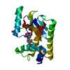
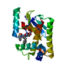
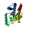
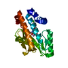
| Title | The N-terminal domain of PA endonuclease from the influenza H1N1 virus in complex with 6-(3-(1H-tetrazol-5-yl)phenyl)-3-hydroxy-4-oxo-1,4-dihydropyridine-2-carboxylic acid | ||||||
 Components Components | Polymerase acidic protein | ||||||
 Keywords Keywords | HYDROLASE/HYDROLASE INHIBITOR / PA endonuclease /  inhibitor / influenza / H1N1 / HYDROLASE-HYDROLASE INHIBITOR complex inhibitor / influenza / H1N1 / HYDROLASE-HYDROLASE INHIBITOR complex | ||||||
| Function / homology |  Function and homology information Function and homology informationsymbiont-mediated suppression of host mRNA transcription via inhibition of RNA polymerase II activity / cap snatching / endonuclease activity / host cell cytoplasm / Hydrolases; Acting on ester bonds / viral RNA genome replication / DNA-templated transcription / host cell nucleus / RNA binding / metal ion bindingSimilarity search - Function | ||||||
| Biological species |   Influenza A virus Influenza A virus | ||||||
| Method | X-RAY DIFFRACTION / MOLECULAR REPLACEMENT / Resolution: 2.8 Å | ||||||
 Authors Authors | Morrison, C.N. / Dick, B.L. / Credille, C.V. / Cohen, S.M. | ||||||
| Funding support |  United States, 1items United States, 1items
| ||||||
 Citation Citation | Journal: J.Med.Chem. / Year: 2019 Title: SAR Exploration of Tight-Binding Inhibitors of Influenza Virus PA Endonuclease. Authors: Credille, C.V. / Morrison, C.N. / Stokes, R.W. / Dick, B.L. / Feng, Y. / Sun, J. / Chen, Y. / Cohen, S.M. | ||||||
| History |
|
- Structure visualization
| Structure viewer | Molecule: MolmilJmol/JSmol |
|---|
- Downloads & links
-Download
| PDBx/mmCIF format | 6e3p.cif.gz | 54.5 KB | Display | PDBx/mmCIF format |
|---|---|---|---|---|
| PDB format | pdb6e3p.ent.gz | 36.9 KB | Display | PDB format |
| PDBx/mmJSON format | 6e3p.json.gz | Tree view | PDBx/mmJSON format | |
| Others |  Other downloads Other downloads |
-Validation report
| Arichive directory | https://data.pdbj.org/pub/pdb/validation_reports/e3/6e3pftp://data.pdbj.org/pub/pdb/validation_reports/e3/6e3p | HTTPS FTP |
|---|
-Related structure data
| Related structure data |  6e3mC  6e3nC  6e3oC  6e4cC  6e6vC 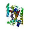 6e6wC  6e6xC  4awmS S: Starting model for refinement C: citing same article ( |
|---|---|
| Similar structure data |


-Links
 PDBj
PDBj- Assembly
| Deposited unit | 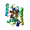
| ||||||||
|---|---|---|---|---|---|---|---|---|---|
| 1 |
| ||||||||
| Unit cell |
|
-Components
| #1: Protein | Mass: 22468.666 Da / Num. of mol.: 1 / Fragment: N-terminal domain (UNP residues 1-198) Source method: isolated from a genetically manipulated source Source: (gene. exp.) Influenza A virus (strain swl A/California/04/2009 H1N1)Strain: swl A/California/04/2009 H1N1 / Gene: PA / Production host:  Escherichia coli BL21 (bacteria) Escherichia coli BL21 (bacteria)References: UniProt: C3W5S0, Hydrolases; Acting on ester bonds | ||||
|---|---|---|---|---|---|
| #2: Chemical |   Mass: 54.938 Da / Num. of mol.: 2 / Source method: obtained synthetically / Formula: Mn Mass: 54.938 Da / Num. of mol.: 2 / Source method: obtained synthetically / Formula: Mn#3: Chemical |   Mass: 299.242 Da / Num. of mol.: 2 / Source method: obtained synthetically / Formula: C13H9N5O4 Mass: 299.242 Da / Num. of mol.: 2 / Source method: obtained synthetically / Formula: C13H9N5O4#4: Water | ChemComp-HOH / | Water Mass: 18.015 Da / Num. of mol.: 5 / Source method: isolated from a natural source / Formula: H2O Mass: 18.015 Da / Num. of mol.: 5 / Source method: isolated from a natural source / Formula: H2O |
-Experimental details
-Experiment
| Experiment | Method: X-RAY DIFFRACTION / Number of used crystals: 1 |
|---|
- Sample preparation
| Crystal | Density Matthews: 2.24 Å3/Da / Density % sol: 45.08 % |
|---|---|
| Crystal grow | Temperature: 306 K / Method: vapor diffusion, hanging drop / pH: 7.8 Details: 30% PEG4000, 100 mM Tris, pH 8.35, 200-220 mM sodium acetate |
-Data collection
| Diffraction | Mean temperature: 100 K |
|---|---|
| Diffraction source | Source: ROTATING ANODE / Type: BRUKER AXS MICROSTAR / Wavelength: 1.54 Å |
| Detector | Type: APEX II CCD / Detector: CCD / Date: Sep 1, 2017 |
| Radiation | Protocol: SINGLE WAVELENGTH / Monochromatic (M) / Laue (L): M / Scattering type: x-ray |
| Radiation wavelength | Wavelength: 1.54 Å / Relative weight: 1 |
| Reflection | Resolution: 2.8→44.25 Å / Num. obs: 5394 / % possible obs: 99.9 % / Redundancy: 14.11 % / Rmerge(I) obs: 0.098 / Net I/σ(I): 20.59 |
| Reflection shell | Resolution: 2.8→2.9 Å / Redundancy: 11.21 % / Rmerge(I) obs: 0.419 / Mean I/σ(I) obs: 3.79 / Num. unique obs: 521 / % possible all: 99.8 |
- Processing
| Software |
| ||||||||||||||||||||||||||||||||||||||||||||||||||||||||||||||||||||||||||||||||||||||||||||||||||||||||||||||||||||||||||||||||||||||||||||||||||||||||||||||||||||||||||||||||||||||
|---|---|---|---|---|---|---|---|---|---|---|---|---|---|---|---|---|---|---|---|---|---|---|---|---|---|---|---|---|---|---|---|---|---|---|---|---|---|---|---|---|---|---|---|---|---|---|---|---|---|---|---|---|---|---|---|---|---|---|---|---|---|---|---|---|---|---|---|---|---|---|---|---|---|---|---|---|---|---|---|---|---|---|---|---|---|---|---|---|---|---|---|---|---|---|---|---|---|---|---|---|---|---|---|---|---|---|---|---|---|---|---|---|---|---|---|---|---|---|---|---|---|---|---|---|---|---|---|---|---|---|---|---|---|---|---|---|---|---|---|---|---|---|---|---|---|---|---|---|---|---|---|---|---|---|---|---|---|---|---|---|---|---|---|---|---|---|---|---|---|---|---|---|---|---|---|---|---|---|---|---|---|---|---|
| Refinement | Method to determine structure: MOLECULAR REPLACEMENT Starting model: 4AWM Resolution: 2.8→44.25 Å / Cor.coef. Fo:Fc: 0.934 / Cor.coef. Fo:Fc free: 0.859 / SU B: 17.349 / SU ML: 0.336 / Cross valid method: THROUGHOUT / ESU R Free: 0.487 / Details: HYDROGENS HAVE BEEN ADDED IN THE RIDING POSITIONS
| ||||||||||||||||||||||||||||||||||||||||||||||||||||||||||||||||||||||||||||||||||||||||||||||||||||||||||||||||||||||||||||||||||||||||||||||||||||||||||||||||||||||||||||||||||||||
| Solvent computation | Ion probe radii: 0.8 Å / Shrinkage radii: 0.8 Å / VDW probe radii: 1.2 Å | ||||||||||||||||||||||||||||||||||||||||||||||||||||||||||||||||||||||||||||||||||||||||||||||||||||||||||||||||||||||||||||||||||||||||||||||||||||||||||||||||||||||||||||||||||||||
| Displacement parameters | Biso mean: 40.261 Å2
| ||||||||||||||||||||||||||||||||||||||||||||||||||||||||||||||||||||||||||||||||||||||||||||||||||||||||||||||||||||||||||||||||||||||||||||||||||||||||||||||||||||||||||||||||||||||
| Refinement step | Cycle: 1 / Resolution: 2.8→44.25 Å
| ||||||||||||||||||||||||||||||||||||||||||||||||||||||||||||||||||||||||||||||||||||||||||||||||||||||||||||||||||||||||||||||||||||||||||||||||||||||||||||||||||||||||||||||||||||||
| Refine LS restraints |
|