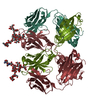
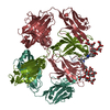
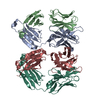
L: anti-HIV-1 Fab 2G12 light chain H: anti-HIV-1 Fab 2G12 heavy chain K: anti-HIV-1 Fab 2G12 light chain M: anti-HIV-1 Fab 2G12 heavy chain hetero molecules
Mass: 23201.840 Da / Num. of mol.: 2 Source method: isolated from a genetically manipulated source Source: (gene. exp.) Homo sapiens (human) / Cell line (production host): 293freestyle / Production host: Homo sapiens (human)
#2: Antibody
anti-HIV-1Fab2G12heavychain
Mass: 23845.791 Da / Num. of mol.: 2 Source method: isolated from a genetically manipulated source Source: (gene. exp.) Homo sapiens (human) / Cell line (production host): 293freestyle / Production host: Homo sapiens (human)
Crystals for 6CXG were grown from a complex of Fab 2G12 and glycopeptide 10F5 ((XXX)SPHLPVLLSK(XXX) ...Crystals for 6CXG were grown from a complex of Fab 2G12 and glycopeptide 10F5 ((XXX)SPHLPVLLSK(XXX)VLNDGRRIVQ(XXX)SSELP(XXX)VRRSGSGSG(BUC)A) where XXX are homopropargylglycine residues covalently linked to Man9 glycans via a cyclohexyl group. authors only see electron density for only the carbohydrate portion of the glycopeptide.
-
Experimental details
-
Experiment
Experiment
Method: X-RAY DIFFRACTION / Number of used crystals: 1
-
Sample preparation
Crystal
Density Matthews: 3.97 Å3/Da / Density % sol: 68.98 %
Crystal grow
Temperature: 295 K / Method: vapor diffusion, sitting drop / pH: 8.5 / Details: 0.2M lithium sulfate, 17.5% Peg400, 0.1M tris
In the structure databanks used in Yorodumi, some data are registered as the other names, "COVID-19 virus" and "2019-nCoV". Here are the details of the virus and the list of structure data.
Jan 31, 2019. EMDB accession codes are about to change! (news from PDBe EMDB page)
EMDB accession codes are about to change! (news from PDBe EMDB page)
The allocation of 4 digits for EMDB accession codes will soon come to an end. Whilst these codes will remain in use, new EMDB accession codes will include an additional digit and will expand incrementally as the available range of codes is exhausted. The current 4-digit format prefixed with “EMD-” (i.e. EMD-XXXX) will advance to a 5-digit format (i.e. EMD-XXXXX), and so on. It is currently estimated that the 4-digit codes will be depleted around Spring 2019, at which point the 5-digit format will come into force.
The EM Navigator/Yorodumi systems omit the EMD- prefix.
Related info.:Q: What is EMD? / ID/Accession-code notation in Yorodumi/EM Navigator
Yorodumi is a browser for structure data from EMDB, PDB, SASBDB, etc.
This page is also the successor to EM Navigator detail page, and also detail information page/front-end page for Omokage search.
The word "yorodu" (or yorozu) is an old Japanese word meaning "ten thousand". "mi" (miru) is to see.
Related info.:EMDB / PDB / SASBDB / Comparison of 3 databanks / Yorodumi Search / Aug 31, 2016. New EM Navigator & Yorodumi / Yorodumi Papers / Jmol/JSmol / Function and homology information / Changes in new EM Navigator and Yorodumi
 Movie
Movie Controller


 Open data
 Basic information
 Components
 Keywords
 IMMUNE SYSTEM /
  Function and homology information
 Authors
 Citation
 Structure visualization
 Downloads & links
 Other downloads
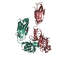


 PDBj
PDBj


 Assembly
 Sample preparation
 / Beamline: BL12-2 / Wavelength: 0.9795 Å
 Processing