 Movie
Movie Controller

[English] 日本語
 Yorodumi
- PDB-6cts: PROPOSED MECHANISM FOR THE CONDENSATION REACTION OF CITRATE SYNTH... -
+ Open data

- Basic information
| Entry | Database: PDB / ID: 6cts | ||||||
|---|---|---|---|---|---|---|---|
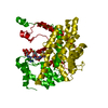
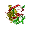
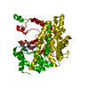
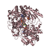
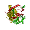
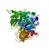
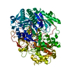
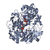
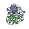
| Title | PROPOSED MECHANISM FOR THE CONDENSATION REACTION OF CITRATE SYNTHASE. 1.9-ANGSTROMS STRUCTURE OF THE TERNARY COMPLEX WITH OXALOACETATE AND CARBOXYMETHYL COENZYME A | ||||||
 Components Components | CITRATE SYNTHASE | ||||||
 Keywords Keywords | OXO-ACID-LYASE | ||||||
| Function / homology |  Function and homology informationcitrate (Si)-synthase / The tricarboxylic acid cycle / citrate (Si)-synthase activity / citrate synthase activity / citrate metabolic process / tricarboxylic acid cycle / carbohydrate metabolic process / mitochondrial matrix / mitochondrion Function and homology informationcitrate (Si)-synthase / The tricarboxylic acid cycle / citrate (Si)-synthase activity / citrate synthase activity / citrate metabolic process / tricarboxylic acid cycle / carbohydrate metabolic process / mitochondrial matrix / mitochondrionSimilarity search - Function | ||||||
| Biological species |  Gallus gallus (chicken) Gallus gallus (chicken) | ||||||
| Method | X-RAY DIFFRACTION / Resolution: 2.5 Å | ||||||
 Authors Authors | Karpusas, M. / Branchaud, B. / Remington, S.J. | ||||||
 Citation Citation | Journal: Biochemistry / Year: 1990 Title: Proposed mechanism for the condensation reaction of citrate synthase: 1.9-A structure of the ternary complex with oxaloacetate and carboxymethyl coenzyme A. Authors: Karpusas, M. / Branchaud, B. / Remington, S.J. #1: Journal: J.Mol.Biol. / Year: 1984Title: Crystal Structure Analysis and Molecular Model of a Complex of Citrate Synthase with Oxaloacetate and S-Acetonyl-Coenzymea Authors: Wiegand, G. / Remington, S. / Deisenhofer, J. / Huber, R. #2: Journal: J.Mol.Biol. / Year: 1982Title: Crystallographic Refinement and Atomic Models of Two Different Forms of Citrate Synthase at 2.7 And 1.7 Angstroms Resolution Authors: Remington, S. / Wiegand, G. / Huber, R. #3: Journal: Proc.Natl.Acad.Sci.USA / Year: 1981Title: Primary Structure of Porcine Heart Citrate Synthase Authors: Bloxham, D.P. / Parmelee, D.C. / Kumar, S. / Wade, R.D. / Ericsson, L.H. / Neurath, H. / Walsh, K.A. / Titani, K. #4: Journal: Eur.J.Biochem. / Year: 1979Title: Crystal Structure Analysis of the Tetragonal Crystal Form and Preliminary Molecular Model of Pig-Heart Citrate Synthase Authors: Wiegand, G. / Kukla, D. / Scholze, H. / Jones, T.A. / Huber, R. | ||||||
| History |
|
- Structure visualization
| Structure viewer | Molecule: MolmilJmol/JSmol |
|---|
- Downloads & links
-Download
| PDBx/mmCIF format | 6cts.cif.gz | 98.1 KB | Display | PDBx/mmCIF format |
|---|---|---|---|---|
| PDB format | pdb6cts.ent.gz | 74.5 KB | Display | PDB format |
| PDBx/mmJSON format | 6cts.json.gz | Tree view | PDBx/mmJSON format | |
| Others |  Other downloads Other downloads |
-Validation report
| Arichive directory | https://data.pdbj.org/pub/pdb/validation_reports/ct/6ctsftp://data.pdbj.org/pub/pdb/validation_reports/ct/6cts | HTTPS FTP |
|---|
-Related structure data


-Links
 PDBj
PDBj

- Assembly
| Deposited unit | 
| ||||||||
|---|---|---|---|---|---|---|---|---|---|
| 1 | 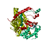
| ||||||||
| Unit cell |
| ||||||||
| Atom site foot note | 1: SEE REMARK 3. | ||||||||
| Details | THE MOLECULE IS A DIMER, WITH ONE MONOMER OF MOLECULAR WEIGHT 50000 IN THE CRYSTALLOGRAPHIC ASYMMETRIC UNIT. THE SYMMETRY-RELATED MONOMER CAN BE GENERATED BY THE FOLLOWING CRYSTALLOGRAPHIC TWO-FOLD OPERATION, XS = - XO YS = YO ZS = - ZO WHERE (XO,YO,ZO) ARE THE COORDINATES IN THIS ENTRY AND (XS,YS,ZS) ARE THE SYMMETRY-RELATED SET. |
-Components
| #1: Protein | Mass: 47322.172 Da / Num. of mol.: 1 Source method: isolated from a genetically manipulated source Source: (gene. exp.) Gallus gallus (chicken) / References: UniProt: P23007, EC: 4.1.3.7 |
|---|---|
| #2: Chemical | ChemComp-CIC /   Mass: 927.659 Da / Num. of mol.: 1 / Source method: obtained synthetically / Formula: C27H44N7O21P3S Mass: 927.659 Da / Num. of mol.: 1 / Source method: obtained synthetically / Formula: C27H44N7O21P3S |
| #3: Water | ChemComp-HOH / Water Mass: 18.015 Da / Num. of mol.: 101 / Source method: isolated from a natural source / Formula: H2O Mass: 18.015 Da / Num. of mol.: 101 / Source method: isolated from a natural source / Formula: H2O |
-Experimental details
-Experiment
| Experiment | Method: X-RAY DIFFRACTION |
|---|
- Sample preparation
| Crystal | Density Matthews: 2.45 Å3/Da / Density % sol: 49.87 % | |||||||||||||||||||||||||
|---|---|---|---|---|---|---|---|---|---|---|---|---|---|---|---|---|---|---|---|---|---|---|---|---|---|---|
| Crystal grow | *PLUS pH: 6 / Method: vapor diffusion / Details: took Remington et al., 1982 from original paper | |||||||||||||||||||||||||
| Components of the solutions | *PLUS
|
-Data collection
| Reflection | *PLUS Highest resolution: 1.9 Å / Num. obs: 30444 / Num. measured all: 61965 |
|---|
- Processing
| Software | Name: TNT / Classification: refinement | ||||||||||||||||||||||||||||||
|---|---|---|---|---|---|---|---|---|---|---|---|---|---|---|---|---|---|---|---|---|---|---|---|---|---|---|---|---|---|---|---|
| Refinement | Resolution: 2.5→6 Å / Rfactor obs: 0.11 | ||||||||||||||||||||||||||||||
| Refinement step | Cycle: LAST / Resolution: 2.5→6 Å
| ||||||||||||||||||||||||||||||
| Refine LS restraints |
| ||||||||||||||||||||||||||||||
| Software | *PLUS Name: TNT / Classification: refinement | ||||||||||||||||||||||||||||||
| Refinement | *PLUS Rfactor obs: 0.11 | ||||||||||||||||||||||||||||||
| Solvent computation | *PLUS | ||||||||||||||||||||||||||||||
| Displacement parameters | *PLUS | ||||||||||||||||||||||||||||||
| Refine LS restraints | *PLUS Type: t_angle_d / Dev ideal: 3.2 |
