 Movie
Movie Controller


+ Open data

- Basic information
| Entry | Database: PDB / ID: 6cs9 | ||||||
|---|---|---|---|---|---|---|---|
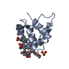
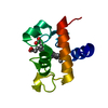
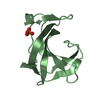
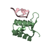
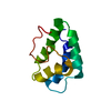
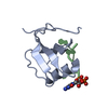
| Title | Crystal structure of human beta-defensin 2 in complex with PIP2 | ||||||
 Components Components | Beta-defensin 4A | ||||||
 Keywords Keywords |  ANTIMICROBIAL PROTEIN / antimicrobial / antifungal / defensin / innate defense ANTIMICROBIAL PROTEIN / antimicrobial / antifungal / defensin / innate defense | ||||||
| Function / homology |  Function and homology information Function and homology informationCCR6 chemokine receptor binding / Beta defensins / Defensins / antifungal innate immune response / chemoattractant activity / defense response to fungus / phosphatidylinositol-4,5-bisphosphate binding / cell chemotaxis / Golgi lumen / chemotaxis ...CCR6 chemokine receptor binding / Beta defensins / Defensins / antifungal innate immune response / chemoattractant activity / defense response to fungus / phosphatidylinositol-4,5-bisphosphate binding / cell chemotaxis / Golgi lumen / chemotaxis / antimicrobial humoral immune response mediated by antimicrobial peptide / killing of cells of another organism / defense response to Gram-negative bacterium / defense response to Gram-positive bacterium / defense response to bacterium / immune response / G protein-coupled receptor signaling pathway / extracellular space / extracellular regionSimilarity search - Function | ||||||
| Biological species |  Homo sapiens (human) Homo sapiens (human) | ||||||
| Method | X-RAY DIFFRACTION / SYNCHROTRON / MOLECULAR REPLACEMENT / molecular replacement / Resolution: 1.85 Å | ||||||
 Authors Authors | Jarva, M. / Phan, K. / Lay, F.T. / Humble, C. / Hulett, M. / Kvansakul, M. | ||||||
| Funding support |  Australia, 1items Australia, 1items
| ||||||
 Citation Citation | Journal: Sci Adv / Year: 2018 Title: Human beta-defensin 2 killsCandida albicansthrough phosphatidylinositol 4,5-bisphosphate-mediated membrane permeabilization. Authors: Jarva, M. / Phan, T.K. / Lay, F.T. / Caria, S. / Kvansakul, M. / Hulett, M.D. | ||||||
| History |
|
- Structure visualization
| Structure viewer | Molecule: MolmilJmol/JSmol |
|---|
- Downloads & links
-Download
| PDBx/mmCIF format | 6cs9.cif.gz | 32.4 KB | Display | PDBx/mmCIF format |
|---|---|---|---|---|
| PDB format | pdb6cs9.ent.gz | 20.3 KB | Display | PDB format |
| PDBx/mmJSON format | 6cs9.json.gz | Tree view | PDBx/mmJSON format | |
| Others |  Other downloads Other downloads |
-Validation report
| Arichive directory | https://data.pdbj.org/pub/pdb/validation_reports/cs/6cs9ftp://data.pdbj.org/pub/pdb/validation_reports/cs/6cs9 | HTTPS FTP |
|---|
-Related structure data
| Related structure data |  1fd4S S: Starting model for refinement |
|---|---|
| Similar structure data | |
| Experimental dataset #1 | Data reference: 10.15785/SBGRID/568 / Data set type: diffraction image data |


-Links
 PDBj
PDBj

- Assembly
| Deposited unit | 
| ||||||||
|---|---|---|---|---|---|---|---|---|---|
| 1 |
| ||||||||
| Unit cell |
|
-Components
| #1: Protein/peptide | Mass: 4342.271 Da / Num. of mol.: 2 Source method: isolated from a genetically manipulated source Source: (gene. exp.) Homo sapiens (human) / Gene: DEFB4A, DEFB102, DEFB2, DEFB4, DEFB4B / Production host:  Komagataella pastoris (fungus) / References: UniProt: O15263 Komagataella pastoris (fungus) / References: UniProt: O15263#2: Chemical |   Mass: 746.566 Da / Num. of mol.: 2 / Source method: obtained synthetically / Formula: C25H49O19P3 / Feature type: SUBJECT OF INVESTIGATION Mass: 746.566 Da / Num. of mol.: 2 / Source method: obtained synthetically / Formula: C25H49O19P3 / Feature type: SUBJECT OF INVESTIGATION#3: Water | ChemComp-HOH / | Water Mass: 18.015 Da / Num. of mol.: 28 / Source method: isolated from a natural source / Formula: H2O Mass: 18.015 Da / Num. of mol.: 28 / Source method: isolated from a natural source / Formula: H2O |
|---|
-Experimental details
-Experiment
| Experiment | Method: X-RAY DIFFRACTION / Number of used crystals: 1 |
|---|
- Sample preparation
| Crystal | Density Matthews: 1.92 Å3/Da / Density % sol: 35.92 % |
|---|---|
| Crystal grow | Temperature: 293.15 K / Method: vapor diffusion, sitting drop / pH: 8.38 Details: 0.697M Sodium-malonate-malonic acid, 0.1M glycine-glycine, pH 8.38 |
-Data collection
| Diffraction | Mean temperature: 100 K | ||||||||||||||||||||||||||||||
|---|---|---|---|---|---|---|---|---|---|---|---|---|---|---|---|---|---|---|---|---|---|---|---|---|---|---|---|---|---|---|---|
| Diffraction source | Source: SYNCHROTRON / Site: Australian Synchrotron / Beamline: MX2 / Wavelength: 0.9537 Å | ||||||||||||||||||||||||||||||
| Detector | Type: ADSC QUANTUM 210r / Detector: CCD / Date: Jul 20, 2016 | ||||||||||||||||||||||||||||||
| Radiation | Protocol: SINGLE WAVELENGTH / Monochromatic (M) / Laue (L): M / Scattering type: x-ray | ||||||||||||||||||||||||||||||
| Radiation wavelength | Wavelength: 0.9537 Å / Relative weight: 1 | ||||||||||||||||||||||||||||||
| Reflection | Resolution: 1.85→39.71 Å / Num. obs: 5703 / % possible obs: 98.4 % / Redundancy: 3.5 % / Biso Wilson estimate: 28.91 Å2 / CC1/2: 0.997 / Rmerge(I) obs: 0.068 / Rpim(I) all: 0.043 / Rrim(I) all: 0.081 / Net I/σ(I): 10.5 / Num. measured all: 20208 | ||||||||||||||||||||||||||||||
| Reflection shell | Diffraction-ID: 1
|
-Phasing
| Phasing | Method: molecular replacement | |||||||||
|---|---|---|---|---|---|---|---|---|---|---|
| Phasing MR |
|
- Processing
| Software |
| ||||||||||||||||||||||||
|---|---|---|---|---|---|---|---|---|---|---|---|---|---|---|---|---|---|---|---|---|---|---|---|---|---|
| Refinement | Method to determine structure: MOLECULAR REPLACEMENT Starting model: 1FD4 Resolution: 1.85→39.714 Å / SU ML: 0.14 / Cross valid method: THROUGHOUT / σ(F): 1.35 / Phase error: 18.89 / Stereochemistry target values: ML
| ||||||||||||||||||||||||
| Solvent computation | Shrinkage radii: 0.9 Å / VDW probe radii: 1.11 Å / Solvent model: FLAT BULK SOLVENT MODEL | ||||||||||||||||||||||||
| Displacement parameters | Biso max: 83.57 Å2 / Biso mean: 36.2562 Å2 / Biso min: 16.14 Å2 | ||||||||||||||||||||||||
| Refinement step | Cycle: final / Resolution: 1.85→39.714 Å
| ||||||||||||||||||||||||
| LS refinement shell | Refine-ID: X-RAY DIFFRACTION / Rfactor Rfree error: 0 / Total num. of bins used: 2
|
