 Movie
Movie Controller

[English] 日本語
 Yorodumi
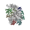
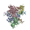
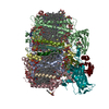
- PDB-6c4h: Conformation of methylated GGQ in the peptidyl transferase center... -
+ Open data

- Basic information
| Entry | Database: PDB / ID: 6c4h | |||||||||
|---|---|---|---|---|---|---|---|---|---|---|
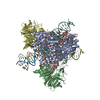
| Title | Conformation of methylated GGQ in the peptidyl transferase center during translation termination (PTC region) | |||||||||
 Components Components |
| |||||||||
 Keywords Keywords |  RIBOSOMAL PROTEIN/RNA / nonstop / termination / ArfA / RF2 / methylation / RIBOSOMAL PROTEIN-RNA complex RIBOSOMAL PROTEIN/RNA / nonstop / termination / ArfA / RF2 / methylation / RIBOSOMAL PROTEIN-RNA complex | |||||||||
| Function / homology |  Function and homology information Function and homology informationtranslation release factor activity, codon specific / positive regulation of ribosome biogenesis / translational termination / DnaA-L2 complex / negative regulation of DNA-templated DNA replication initiation / assembly of large subunit precursor of preribosome / cytosolic ribosome assembly / regulation of cell growth / ribosomal large subunit assembly / ribosome binding ...translation release factor activity, codon specific / positive regulation of ribosome biogenesis / translational termination / DnaA-L2 complex / negative regulation of DNA-templated DNA replication initiation / assembly of large subunit precursor of preribosome / cytosolic ribosome assembly / regulation of cell growth / ribosomal large subunit assembly / ribosome binding / cytoplasmic translation / cytosolic large ribosomal subunit / transferase activity / tRNA binding / rRNA binding / structural constituent of ribosome / RNA binding / zinc ion binding / cytosol / cytoplasmSimilarity search - Function | |||||||||
| Biological species |  Escherichia coli (E. coli) Escherichia coli (E. coli) | |||||||||
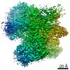
| Method | ELECTRON MICROSCOPY / single particle reconstruction / cryo EM / Resolution: 3.1 Å | |||||||||
 Authors Authors | Zeng, F. / Jin, H. | |||||||||
| Funding support |  United States, 1items United States, 1items
| |||||||||
 Citation Citation | Journal: Sci Rep / Year: 2018 Title: Conformation of methylated GGQ in the Peptidyl Transferase Center during Translation Termination. Authors: Fuxing Zeng / Hong Jin / Abstract: The universally conserved Gly-Gly-Gln (GGQ) tripeptide in release factors or release factor-like surveillance proteins is required to catalyze the release of nascent peptide in the ribosome. The ...The universally conserved Gly-Gly-Gln (GGQ) tripeptide in release factors or release factor-like surveillance proteins is required to catalyze the release of nascent peptide in the ribosome. The glutamine of the GGQ is methylated post-translationally at the N position in vivo; this covalent modification is essential for optimal cell growth and efficient translation termination. However, the precise conformation of the methylated-GGQ tripeptide in the ribosome remains unknown. Using cryoEM and X-ray crystallography, we report the conformation of methylated-GGQ in the peptidyl transferase center of the ribosome during canonical translational termination and co-translation quality control. It has been suggested that the GGQ motif arose independently through convergent evolution among otherwise unrelated proteins that catalyze peptide release. The requirement for this tripeptide in the highly conserved peptidyl transferase center suggests that the conformation reported here is likely shared during termination of protein synthesis in all domains of life. | |||||||||
| History |
|
- Structure visualization
| Movie |
Movie viewer |
|---|---|
| Structure viewer | Molecule: MolmilJmol/JSmol |
- Downloads & links
-Download
| PDBx/mmCIF format | 6c4h.cif.gz | 445.3 KB | Display | PDBx/mmCIF format |
|---|---|---|---|---|
| PDB format | pdb6c4h.ent.gz | 319.9 KB | Display | PDB format |
| PDBx/mmJSON format | 6c4h.json.gz | Tree view | PDBx/mmJSON format | |
| Others |  Other downloads Other downloads |
-Validation report
| Arichive directory | https://data.pdbj.org/pub/pdb/validation_reports/c4/6c4hftp://data.pdbj.org/pub/pdb/validation_reports/c4/6c4h | HTTPS FTP |
|---|
-Related structure data
| Related structure data | 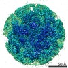 7340MC 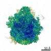 7341C  6c4iC 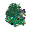 6c5lC M: map data used to model this data C: citing same article ( |
|---|---|
| Similar structure data |

-Links
 PDBj
PDBj


- Assembly
| Deposited unit | 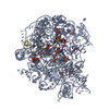
|
|---|---|
| 1 |
|
-Components
-RNA chain , 2 types, 2 molecules Ax
| #1: RNA chain | 23S ribosomal RNA Mass: 941818.625 Da / Num. of mol.: 1 / Source method: isolated from a natural source / Source: (natural) Escherichia coli (E. coli) / References: GenBank: 687670942 |
|---|---|
| #6: RNA chain | Mass: 24786.785 Da / Num. of mol.: 1 Source method: isolated from a genetically manipulated source Source: (gene. exp.) Escherichia coli (E. coli) / Production host: Escherichia coli (E. coli) / References: GenBank: 817573384 |
-50S ribosomal protein ... , 4 types, 4 molecules CDNX
| #2: Protein | / Large ribosomal subunit protein uL2 Mass: 29923.619 Da / Num. of mol.: 1 / Source method: isolated from a natural source / Source: (natural) Escherichia coli (E. coli) / References: UniProt: P60422 |
|---|---|
| #3: Protein | / Large ribosomal subunit protein uL3 Mass: 22277.535 Da / Num. of mol.: 1 / Source method: isolated from a natural source / Source: (natural) Escherichia coli (E. coli) / References: UniProt: P60438 |
| #4: Protein | / Large ribosomal subunit protein uL16 Mass: 15312.269 Da / Num. of mol.: 1 / Source method: isolated from a natural source / Source: (natural) Escherichia coli (E. coli) / References: UniProt: P0ADY7 |
| #5: Protein | / Large ribosomal subunit protein bL27 Mass: 9146.540 Da / Num. of mol.: 1 / Source method: isolated from a natural source / Source: (natural) Escherichia coli (E. coli) / References: UniProt: P0A7L8 |
-Protein / Non-polymers , 2 types, 35 molecules v
| #7: Protein | Mass: 43384.945 Da / Num. of mol.: 1 Source method: isolated from a genetically manipulated source Source: (gene. exp.) Escherichia coli (E. coli) / Gene: prfB, supK, b2891, JW5847 / Production host: Escherichia coli (E. coli) / References: UniProt: P07012 |
|---|---|
| #8: Chemical | ChemComp-MG /  Mass: 24.305 Da / Num. of mol.: 34 / Source method: obtained synthetically / Formula: Mg Mass: 24.305 Da / Num. of mol.: 34 / Source method: obtained synthetically / Formula: Mg |
-Experimental details
-Experiment
| Experiment | Method: ELECTRON MICROSCOPY |
|---|---|
| EM experiment | Aggregation state: PARTICLE / 3D reconstruction method: single particle reconstruction |
- Sample preparation
| Component |
| ||||||||||||||||||||||||||||||||||||||||||||||||||||||||||||||||||||||
|---|---|---|---|---|---|---|---|---|---|---|---|---|---|---|---|---|---|---|---|---|---|---|---|---|---|---|---|---|---|---|---|---|---|---|---|---|---|---|---|---|---|---|---|---|---|---|---|---|---|---|---|---|---|---|---|---|---|---|---|---|---|---|---|---|---|---|---|---|---|---|---|
| Source (natural) |
| ||||||||||||||||||||||||||||||||||||||||||||||||||||||||||||||||||||||
| Source (recombinant) |
| ||||||||||||||||||||||||||||||||||||||||||||||||||||||||||||||||||||||
| Buffer solution | pH: 7.5 | ||||||||||||||||||||||||||||||||||||||||||||||||||||||||||||||||||||||
| Buffer component |
| ||||||||||||||||||||||||||||||||||||||||||||||||||||||||||||||||||||||
| Specimen | Embedding applied: NO / Shadowing applied: NO / Staining applied: NO / Vitrification applied: YES | ||||||||||||||||||||||||||||||||||||||||||||||||||||||||||||||||||||||
| Specimen support | Grid type: C-flat-2/0.5 4C | ||||||||||||||||||||||||||||||||||||||||||||||||||||||||||||||||||||||
| Vitrification | Instrument: FEI VITROBOT MARK IV / Cryogen name: ETHANE / Humidity: 100 % / Chamber temperature: 277 K / Details: Grids were blotted for 3.5 seconds. |
- Electron microscopy imaging
| Microscopy | Model: JEOL 3200FS |
|---|---|
| Electron gun | Electron source: FIELD EMISSION GUN / Accelerating voltage: 300 kV / Illumination mode: FLOOD BEAM |
| Electron lens | Mode: BRIGHT FIELDBright-field microscopy / Calibrated magnification: 83822 X |
| Specimen holder | Cryogen: NITROGEN |
| Image recording | Electron dose: 20 e/Å2 / Detector mode: SUPER-RESOLUTION / Film or detector model: GATAN K2 SUMMIT (4k x 4k) |
- Processing
| EM software |
| ||||||||||||||||||||||||||||||||||||||||||||
|---|---|---|---|---|---|---|---|---|---|---|---|---|---|---|---|---|---|---|---|---|---|---|---|---|---|---|---|---|---|---|---|---|---|---|---|---|---|---|---|---|---|---|---|---|---|
| CTF correction | Type: PHASE FLIPPING AND AMPLITUDE CORRECTION | ||||||||||||||||||||||||||||||||||||||||||||
| Symmetry | Point symmetry: C1 (asymmetric) | ||||||||||||||||||||||||||||||||||||||||||||
| 3D reconstruction | Resolution: 3.1 Å / Resolution method: FSC 0.143 CUT-OFF / Num. of particles: 143372 / Num. of class averages: 1 / Symmetry type: POINT | ||||||||||||||||||||||||||||||||||||||||||||
| Atomic model building | Protocol: FLEXIBLE FIT / Space: RECIPROCAL / Target criteria: Average FSC |
