 Movie
Movie Controller

[English] 日本語
 Yorodumi
- PDB-6aoy: F9 pilus adhesin FmlH lectin domain from E. coli UTI89 co-crystal... -
+ Open data

- Basic information
| Entry | Database: PDB / ID: 6aoy | |||||||||
|---|---|---|---|---|---|---|---|---|---|---|
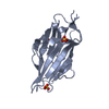
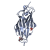
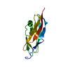
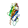
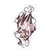
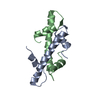
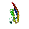
| Title | F9 pilus adhesin FmlH lectin domain from E. coli UTI89 co-crystallized with o-nitrophenyl beta-galactoside (ONPG) | |||||||||
 Components Components | Fml fimbrial adhesin FmlD | |||||||||
 Keywords Keywords | SUGAR BINDING PROTEIN / Fimbrial adhesin /  lectin / bacterial adhesion lectin / bacterial adhesion | |||||||||
| Function / homology |  Function and homology information Function and homology information | |||||||||
| Biological species |  Escherichia coli (E. coli) Escherichia coli (E. coli) | |||||||||
| Method | X-RAY DIFFRACTION / MOLECULAR REPLACEMENT / Resolution: 1.8 Å | |||||||||
 Authors Authors | Kalas, V. / Hultgren, S.J. | |||||||||
 Citation Citation | Journal: Proc.Natl.Acad.Sci.USA / Year: 2018 Title: Structure-based discovery of glycomimetic FmlH ligands as inhibitors of bacterial adhesion during urinary tract infection. Authors: Kalas, V. / Hibbing, M.E. / Maddirala, A.R. / Chugani, R. / Pinkner, J.S. / Mydock-McGrane, L.K. / Conover, M.S. / Janetka, J.W. / Hultgren, S.J. | |||||||||
| History |
|
- Structure visualization
| Structure viewer | Molecule: MolmilJmol/JSmol |
|---|
- Downloads & links
-Download
| PDBx/mmCIF format | 6aoy.cif.gz | 80.7 KB | Display | PDBx/mmCIF format |
|---|---|---|---|---|
| PDB format | pdb6aoy.ent.gz | 58.2 KB | Display | PDB format |
| PDBx/mmJSON format | 6aoy.json.gz | Tree view | PDBx/mmJSON format | |
| Others |  Other downloads Other downloads |
-Validation report
| Arichive directory | https://data.pdbj.org/pub/pdb/validation_reports/ao/6aoyftp://data.pdbj.org/pub/pdb/validation_reports/ao/6aoy | HTTPS FTP |
|---|
-Related structure data
| Related structure data |  6aowSC  6aoxC 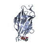 6armC 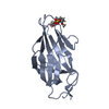 6arnC 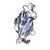 6aroC 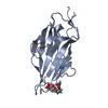 6as8C S: Starting model for refinement C: citing same article ( |
|---|---|
| Similar structure data |


-Links
 PDBj
PDBj- Assembly
| Deposited unit | 
| |||||||||||||||
|---|---|---|---|---|---|---|---|---|---|---|---|---|---|---|---|---|
| 1 | 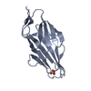
| |||||||||||||||
| 2 | 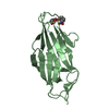
| |||||||||||||||
| Unit cell |
| |||||||||||||||
| Components on special symmetry positions |
|
-Components
| #1: Protein | Mass: 17920.088 Da / Num. of mol.: 2 Source method: isolated from a genetically manipulated source Source: (gene. exp.) Escherichia coli (E. coli) / Strain: UTI89 / UPEC / Gene: fmlD, UTI89_C1716 / Production host: Escherichia coli (E. coli) / Strain (production host): C600 / References: UniProt: Q1RBS0#2: Chemical | ChemComp-SO4 / | Sulfate  Mass: 96.063 Da / Num. of mol.: 1 / Source method: obtained synthetically / Formula: SO4 Mass: 96.063 Da / Num. of mol.: 1 / Source method: obtained synthetically / Formula: SO4#3: Sugar | ChemComp-145 / |   Type: D-saccharide / Mass: 301.249 Da / Num. of mol.: 1 / Source method: obtained synthetically / Formula: C12H15NO8 / Feature type: SUBJECT OF INVESTIGATION Type: D-saccharide / Mass: 301.249 Da / Num. of mol.: 1 / Source method: obtained synthetically / Formula: C12H15NO8 / Feature type: SUBJECT OF INVESTIGATION#4: Water | ChemComp-HOH / | Water Mass: 18.015 Da / Num. of mol.: 263 / Source method: isolated from a natural source / Formula: H2O Mass: 18.015 Da / Num. of mol.: 263 / Source method: isolated from a natural source / Formula: H2O |
|---|
-Experimental details
-Experiment
| Experiment | Method: X-RAY DIFFRACTION / Number of used crystals: 1 |
|---|
- Sample preparation
| Crystal | Density Matthews: 2.1 Å3/Da / Density % sol: 41.41 % |
|---|---|
| Crystal grow | Temperature: 293 K / Method: vapor diffusion, hanging drop / pH: 5.6 Details: 0.2 M ammonium sulfate, 0.1M NaCl, 0.1 M MES pH 5.6, 32% PEG 3350 |
-Data collection
| Diffraction | Mean temperature: 100 K |
|---|---|
| Diffraction source | Source: ROTATING ANODE / Type: RIGAKU MICROMAX-007 / Wavelength: 1.5418 Å |
| Detector | Type: MAR scanner 345 mm plate / Detector: IMAGE PLATE / Date: Nov 24, 2014 |
| Radiation | Protocol: SINGLE WAVELENGTH / Monochromatic (M) / Laue (L): M / Scattering type: x-ray |
| Radiation wavelength | Wavelength: 1.5418 Å / Relative weight: 1 |
| Reflection | Resolution: 1.8→40.03 Å / Num. obs: 27380 / % possible obs: 99.6 % / Redundancy: 6.9 % / CC1/2: 0.999 / Rmerge(I) obs: 0.081 / Rpim(I) all: 0.032 / Rrim(I) all: 0.087 / Net I/σ(I): 15.6 |
| Reflection shell | Resolution: 1.8→1.9 Å / Redundancy: 5.1 % / Rmerge(I) obs: 0.678 / Mean I/σ(I) obs: 2.8 / Num. unique obs: 3937 / CC1/2: 0.788 / Rpim(I) all: 0.322 / Rrim(I) all: 0.754 / % possible all: 98.5 |
- Processing
| Software |
| |||||||||||||||||||||||||||||||||||||||||||||||||||||||||||||||||||||||||||||
|---|---|---|---|---|---|---|---|---|---|---|---|---|---|---|---|---|---|---|---|---|---|---|---|---|---|---|---|---|---|---|---|---|---|---|---|---|---|---|---|---|---|---|---|---|---|---|---|---|---|---|---|---|---|---|---|---|---|---|---|---|---|---|---|---|---|---|---|---|---|---|---|---|---|---|---|---|---|---|
| Refinement | Method to determine structure: MOLECULAR REPLACEMENT Starting model: 6AOW Resolution: 1.8→32.83 Å / SU ML: 0.21 / Cross valid method: FREE R-VALUE / σ(F): 1.36 / Phase error: 22.84
| |||||||||||||||||||||||||||||||||||||||||||||||||||||||||||||||||||||||||||||
| Solvent computation | Shrinkage radii: 0.9 Å / VDW probe radii: 1.11 Å | |||||||||||||||||||||||||||||||||||||||||||||||||||||||||||||||||||||||||||||
| Refinement step | Cycle: LAST / Resolution: 1.8→32.83 Å
| |||||||||||||||||||||||||||||||||||||||||||||||||||||||||||||||||||||||||||||
| Refine LS restraints |
| |||||||||||||||||||||||||||||||||||||||||||||||||||||||||||||||||||||||||||||
| LS refinement shell |
|
