 Movie
Movie Controller


+ Open data

- Basic information
| Entry | Database: PDB / ID: 6akk | ||||||
|---|---|---|---|---|---|---|---|
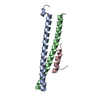
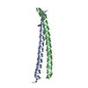
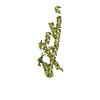
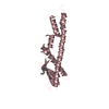
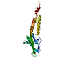
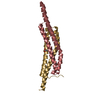
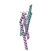
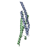
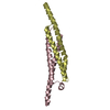
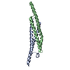
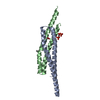
| Title | Crystal structure of the second Coiled-coil domain of SIKE1 | ||||||
 Components Components | Suppressor of IKBKE 1 | ||||||
 Keywords Keywords |  PROTEIN BINDING / homotetramer / Coiled-coil PROTEIN BINDING / homotetramer / Coiled-coil | ||||||
| Function / homology |  Function and homology information Function and homology informationTRAF6 mediated IRF7 activation / small GTPase binding / SARS-CoV-1 activates/modulates innate immune responses / TRAF3-dependent IRF activation pathway / protein kinase binding / SARS-CoV-2 activates/modulates innate and adaptive immune responses / cytosolSimilarity search - Function | ||||||
| Biological species |  Homo sapiens (human) Homo sapiens (human) | ||||||
| Method | X-RAY DIFFRACTION / SYNCHROTRON / SAD / Resolution: 1.5 Å | ||||||
 Authors Authors | Zhou, L. / Chen, M. / Zhou, Z.C. | ||||||
 Citation Citation | Journal: Cell Discov / Year: 2019 Title: Architecture, substructures, and dynamic assembly of STRIPAK complexes in Hippo signaling. Authors: Tang, Y. / Chen, M. / Zhou, L. / Ma, J. / Li, Y. / Zhang, H. / Shi, Z. / Xu, Q. / Zhang, X. / Gao, Z. / Zhao, Y. / Cheng, Y. / Jiao, S. / Zhou, Z. | ||||||
| History |
|
- Structure visualization
| Structure viewer | Molecule: MolmilJmol/JSmol |
|---|
- Downloads & links
-Download
| PDBx/mmCIF format | 6akk.cif.gz | 55.8 KB | Display | PDBx/mmCIF format |
|---|---|---|---|---|
| PDB format | pdb6akk.ent.gz | 40.3 KB | Display | PDB format |
| PDBx/mmJSON format | 6akk.json.gz | Tree view | PDBx/mmJSON format | |
| Others |  Other downloads Other downloads |
-Validation report
| Arichive directory | https://data.pdbj.org/pub/pdb/validation_reports/ak/6akkftp://data.pdbj.org/pub/pdb/validation_reports/ak/6akk | HTTPS FTP |
|---|
-Related structure data

-Links
 PDBj
PDBj

- Assembly
| Deposited unit | 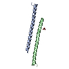
| ||||||||
|---|---|---|---|---|---|---|---|---|---|
| 1 | 
| ||||||||
| Unit cell |
| ||||||||
| Components on special symmetry positions |
|
-Components
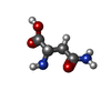
| #1: Protein | Mass: 6426.421 Da / Num. of mol.: 2 Source method: isolated from a genetically manipulated source Source: (gene. exp.) Homo sapiens (human) / Gene: SIKE1, SIKE / Production host:  Escherichia coli BL21(DE3) (bacteria) / Strain (production host): BL21 (DE3) / Variant (production host): CodonPlus / References: UniProt: Q9BRV8 Escherichia coli BL21(DE3) (bacteria) / Strain (production host): BL21 (DE3) / Variant (production host): CodonPlus / References: UniProt: Q9BRV8#2: Chemical | ChemComp-GOL / | Glycerol  Mass: 92.094 Da / Num. of mol.: 1 / Source method: isolated from a natural source / Formula: C3H8O3 Mass: 92.094 Da / Num. of mol.: 1 / Source method: isolated from a natural source / Formula: C3H8O3#3: Water | ChemComp-HOH / | Water Mass: 18.015 Da / Num. of mol.: 99 / Source method: isolated from a natural source / Formula: H2O Mass: 18.015 Da / Num. of mol.: 99 / Source method: isolated from a natural source / Formula: H2O |
|---|
-Experimental details
-Experiment
| Experiment | Method: X-RAY DIFFRACTION / Number of used crystals: 1 |
|---|
- Sample preparation
| Crystal | Density Matthews: 2.14 Å3/Da / Density % sol: 42.48 % |
|---|---|
| Crystal grow | Temperature: 289 K / Method: vapor diffusion, sitting drop Details: 1.0 M (NH4)2HPO4 (ammonium dibasic phosphate); 0.1 M acetate pH 4.5 |
-Data collection
| Diffraction | Mean temperature: 100 K / Serial crystal experiment: N |
|---|---|
| Diffraction source | Source: SYNCHROTRON / Site: SSRF  / Beamline: BL17U1 / Wavelength: 0.97915 Å / Beamline: BL17U1 / Wavelength: 0.97915 Å |
| Detector | Type: ADSC QUANTUM 315 / Detector: CCD / Date: May 27, 2017 |
| Radiation | Protocol: SINGLE WAVELENGTH / Monochromatic (M) / Laue (L): M / Scattering type: x-ray |
| Radiation wavelength | Wavelength: 0.97915 Å / Relative weight: 1 |
| Reflection | Resolution: 1.5→50 Å / Num. obs: 17636 / % possible obs: 97.6 % / Redundancy: 13.4 % / Rmerge(I) obs: 0.091 / Rpim(I) all: 0.026 / Rrim(I) all: 0.095 / Net I/σ(I): 32.5 |
| Reflection shell | Resolution: 1.5→1.53 Å / Redundancy: 14.1 % / Rmerge(I) obs: 0.582 / Mean I/σ(I) obs: 5.1 / Num. unique obs: 873 / CC1/2: 0.994 / % possible all: 100 |
- Processing
| Software |
| |||||||||||||||||||||||||||||||||||||||||||||||||||||||||||||||||||||||||||||||||||||||||||
|---|---|---|---|---|---|---|---|---|---|---|---|---|---|---|---|---|---|---|---|---|---|---|---|---|---|---|---|---|---|---|---|---|---|---|---|---|---|---|---|---|---|---|---|---|---|---|---|---|---|---|---|---|---|---|---|---|---|---|---|---|---|---|---|---|---|---|---|---|---|---|---|---|---|---|---|---|---|---|---|---|---|---|---|---|---|---|---|---|---|---|---|---|
| Refinement | Method to determine structure: SAD / Resolution: 1.5→35.598 Å / SU ML: 0.15 / Cross valid method: FREE R-VALUE / σ(F): 0 / Phase error: 29.21 / Stereochemistry target values: ML
| |||||||||||||||||||||||||||||||||||||||||||||||||||||||||||||||||||||||||||||||||||||||||||
| Solvent computation | Shrinkage radii: 0.9 Å / VDW probe radii: 1.11 Å / Solvent model: FLAT BULK SOLVENT MODEL | |||||||||||||||||||||||||||||||||||||||||||||||||||||||||||||||||||||||||||||||||||||||||||
| Refinement step | Cycle: LAST / Resolution: 1.5→35.598 Å
| |||||||||||||||||||||||||||||||||||||||||||||||||||||||||||||||||||||||||||||||||||||||||||
| Refine LS restraints |
| |||||||||||||||||||||||||||||||||||||||||||||||||||||||||||||||||||||||||||||||||||||||||||
| LS refinement shell |
| |||||||||||||||||||||||||||||||||||||||||||||||||||||||||||||||||||||||||||||||||||||||||||
| Refinement TLS params. | Method: refined / Refine-ID: X-RAY DIFFRACTION
| |||||||||||||||||||||||||||||||||||||||||||||||||||||||||||||||||||||||||||||||||||||||||||
| Refinement TLS group |
|
