 Movie
Movie Controller

[English] 日本語
 Yorodumi
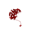
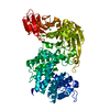
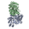
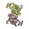
- PDB-6abw: Crystal structure of citrate synthase (Msed_0281) from Metallosph... -
+ Open data

- Basic information
| Entry | Database: PDB / ID: 6abw | ||||||
|---|---|---|---|---|---|---|---|
| Title | Crystal structure of citrate synthase (Msed_0281) from Metallosphaera sedula in complex with acetyl-CoA | ||||||
 Components Components | Citrate synthase | ||||||
 Keywords Keywords | TRANSFERASE / Citrate synthase / Metallosphaera sedula | ||||||
| Function / homology |  Function and homology information Function and homology informationcitrate synthase (unknown stereospecificity) / citrate synthase activity / tricarboxylic acid cycle / membrane / cytoplasmSimilarity search - Function | ||||||
| Biological species |  Metallosphaera sedula (archaea) Metallosphaera sedula (archaea) | ||||||
| Method | X-RAY DIFFRACTION / SYNCHROTRON / MOLECULAR REPLACEMENT / Resolution: 1.72 Å | ||||||
 Authors Authors | Lee, S.-H. / Kim, K.-J. | ||||||
 Citation Citation | Journal: Biochem. Biophys. Res. Commun. / Year: 2019 Title: Crystal structure and biochemical properties of msed_0281, the citrate synthase from Metallosphaera sedula. Authors: Lee, S.H. / Kim, K.J. | ||||||
| History |
|
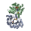
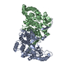
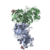
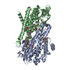
- Structure visualization
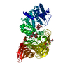
| Structure viewer | Molecule: MolmilJmol/JSmol |
|---|
- Downloads & links
-Download
| PDBx/mmCIF format | 6abw.cif.gz | 95.8 KB | Display | PDBx/mmCIF format |
|---|---|---|---|---|
| PDB format | pdb6abw.ent.gz | 70 KB | Display | PDB format |
| PDBx/mmJSON format | 6abw.json.gz | Tree view | PDBx/mmJSON format | |
| Others |  Other downloads Other downloads |
-Validation report
| Arichive directory | https://data.pdbj.org/pub/pdb/validation_reports/ab/6abwftp://data.pdbj.org/pub/pdb/validation_reports/ab/6abw | HTTPS FTP |
|---|
-Related structure data
| Related structure data |  6abvC  1gvmS S: Starting model for refinement C: citing same article ( |
|---|---|
| Similar structure data |

-Links
 PDBj
PDBj

- Assembly
| Deposited unit | 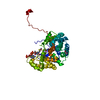
| |||||||||
|---|---|---|---|---|---|---|---|---|---|---|
| 1 | 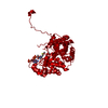
| |||||||||
| Unit cell |
| |||||||||
| Components on special symmetry positions |
|
-Components
| #1: Protein | Mass: 43760.926 Da / Num. of mol.: 1 Source method: isolated from a genetically manipulated source Source: (gene. exp.) Metallosphaera sedula (strain ATCC 51363 / DSM 5348 / JCM 9185 / NBRC 15509 / TH2) (archaea)Strain: ATCC 51363 / DSM 5348 / JCM 9185 / NBRC 15509 / TH2 / Gene: Msed_0281 / Production host:  Escherichia coli (E. coli) Escherichia coli (E. coli)References: UniProt: A4YDF6, citrate synthase (unknown stereospecificity) | ||||
|---|---|---|---|---|---|
| #2: Chemical | Glycerol  Mass: 92.094 Da / Num. of mol.: 2 / Source method: obtained synthetically / Formula: C3H8O3 Mass: 92.094 Da / Num. of mol.: 2 / Source method: obtained synthetically / Formula: C3H8O3#3: Chemical | ChemComp-ACO / | Acetyl-CoA  Mass: 809.571 Da / Num. of mol.: 1 / Source method: obtained synthetically / Formula: C23H38N7O17P3S Mass: 809.571 Da / Num. of mol.: 1 / Source method: obtained synthetically / Formula: C23H38N7O17P3S#4: Water | ChemComp-HOH / | Water Mass: 18.015 Da / Num. of mol.: 244 / Source method: isolated from a natural source / Formula: H2O Mass: 18.015 Da / Num. of mol.: 244 / Source method: isolated from a natural source / Formula: H2O |
-Experimental details
-Experiment
| Experiment | Method: X-RAY DIFFRACTION / Number of used crystals: 1 |
|---|
- Sample preparation
| Crystal | Density Matthews: 2.17 Å3/Da / Density % sol: 43.23 % |
|---|---|
| Crystal grow | Temperature: 295 K / Method: vapor diffusion, sitting drop / pH: 5 Details: 35% v/v (+/-)-2-Methyl-2,4- pentandiol (MPD), 0.1 M Bis-Tris, pH 5.0, 0.2 M ammonium acetate |
-Data collection
| Diffraction | Mean temperature: 100 K |
|---|---|
| Diffraction source | Source: SYNCHROTRON / Site: PAL/PLS  / Beamline: 7A (6B, 6C1) / Wavelength: 0.97934 Å / Beamline: 7A (6B, 6C1) / Wavelength: 0.97934 Å |
| Detector | Type: ADSC QUANTUM 270 / Detector: CCD / Date: May 9, 2017 |
| Radiation | Protocol: SINGLE WAVELENGTH / Monochromatic (M) / Laue (L): M / Scattering type: x-ray |
| Radiation wavelength | Wavelength: 0.97934 Å / Relative weight: 1 |
| Reflection | Resolution: 1.72→64.94 Å / Num. obs: 39803 / % possible obs: 98.1 % / Redundancy: 9.4 % / Rmerge(I) obs: 0.092 / Net I/σ(I): 40.3 |
| Reflection shell | Resolution: 1.72→1.75 Å / Rmerge(I) obs: 0.293 / Num. unique obs: 1928 |
- Processing
| Software |
| ||||||||||||||||||||||||||||||||||||||||||||||||||||||||||||
|---|---|---|---|---|---|---|---|---|---|---|---|---|---|---|---|---|---|---|---|---|---|---|---|---|---|---|---|---|---|---|---|---|---|---|---|---|---|---|---|---|---|---|---|---|---|---|---|---|---|---|---|---|---|---|---|---|---|---|---|---|---|
| Refinement | Method to determine structure: MOLECULAR REPLACEMENT Starting model: 1GVM Resolution: 1.72→64.93 Å / Cor.coef. Fo:Fc: 0.967 / Cor.coef. Fo:Fc free: 0.943 / SU B: 2.164 / SU ML: 0.069 / Cross valid method: THROUGHOUT / σ(F): 0 / ESU R: 0.095 / ESU R Free: 0.101 Details: HYDROGENS HAVE BEEN ADDED IN THE RIDING POSITIONS U VALUES : REFINED INDIVIDUALLY
| ||||||||||||||||||||||||||||||||||||||||||||||||||||||||||||
| Solvent computation | Ion probe radii: 0.8 Å / Shrinkage radii: 0.8 Å / VDW probe radii: 1.2 Å | ||||||||||||||||||||||||||||||||||||||||||||||||||||||||||||
| Displacement parameters | Biso max: 109.41 Å2 / Biso mean: 21.223 Å2 / Biso min: 6.14 Å2
| ||||||||||||||||||||||||||||||||||||||||||||||||||||||||||||
| Refinement step | Cycle: final / Resolution: 1.72→64.93 Å
| ||||||||||||||||||||||||||||||||||||||||||||||||||||||||||||
| Refine LS restraints |
| ||||||||||||||||||||||||||||||||||||||||||||||||||||||||||||
| LS refinement shell | Resolution: 1.723→1.768 Å / Rfactor Rfree error: 0 / Total num. of bins used: 20
|
