 Movie
Movie Controller


+ Open data

- Basic information
| Entry | Database: PDB / ID: 6a9s | |||||||||
|---|---|---|---|---|---|---|---|---|---|---|
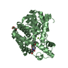
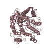
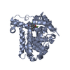
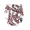
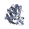
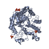
| Title | The crystal structure of vaccinia virus A26 (residues 1-397) | |||||||||
 Components Components | Protein A26 | |||||||||
 Keywords Keywords |  VIRAL PROTEIN / Vaccinia virus / virus entry / acid sensitive VIRAL PROTEIN / Vaccinia virus / virus entry / acid sensitive | |||||||||
| Function / homology | Orthopoxvirus A26L/A30L / Orthopoxvirus A26L/A30L protein / Chordopoxvirus fusion protein/multifunctional envelope protein A27 / Chordopoxvirus multifunctional envelope protein A27 / fusion of virus membrane with host plasma membrane / viral envelope / virion membrane / Protein A26 Function and homology information Function and homology information | |||||||||
| Biological species |  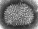 Vaccinia virus Vaccinia virus | |||||||||
| Method | X-RAY DIFFRACTION / SYNCHROTRON / SAD / Resolution: 1.18 Å | |||||||||
 Authors Authors | Wang, H.C. / Ko, T.Z. / Luo, Y.C. / Liao, Y.T. / Chang, W. | |||||||||
| Funding support |  Taiwan, 2items Taiwan, 2items
| |||||||||
 Citation Citation | Journal: Plos Pathog. / Year: 2019 Title: Vaccinia viral A26 protein is a fusion suppressor of mature virus and triggers membrane fusion through conformational change at low pH. Authors: Chang, H.W. / Yang, C.H. / Luo, Y.C. / Su, B.G. / Cheng, H.Y. / Tung, S.Y. / Carillo, K.J.D. / Liao, Y.T. / Tzou, D.M. / Wang, H.C. / Chang, W. | |||||||||
| History |
|
- Structure visualization
| Structure viewer | Molecule: MolmilJmol/JSmol |
|---|
- Downloads & links
-Download
| PDBx/mmCIF format | 6a9s.cif.gz | 183.4 KB | Display | PDBx/mmCIF format |
|---|---|---|---|---|
| PDB format | pdb6a9s.ent.gz | 151.4 KB | Display | PDB format |
| PDBx/mmJSON format | 6a9s.json.gz | Tree view | PDBx/mmJSON format | |
| Others |  Other downloads Other downloads |
-Validation report
| Arichive directory | https://data.pdbj.org/pub/pdb/validation_reports/a9/6a9sftp://data.pdbj.org/pub/pdb/validation_reports/a9/6a9s | HTTPS FTP |
|---|
-Related structure data
| Similar structure data |
|---|


-Links
 PDBj
PDBj- Assembly
| Deposited unit | 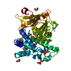
| ||||||||
|---|---|---|---|---|---|---|---|---|---|
| 1 |
| ||||||||
| Unit cell |
|
-Components
| #1: Protein | Mass: 46791.219 Da / Num. of mol.: 1 Source method: isolated from a genetically manipulated source Source: (gene. exp.) Vaccinia virus (strain Western Reserve)Strain: Western Reserve / Gene: VACWR149, A26L / Production host:  Escherichia coli (E. coli) / References: UniProt: P24758 Escherichia coli (E. coli) / References: UniProt: P24758 | ||
|---|---|---|---|
| #2: Chemical | ChemComp-EDO / Ethylene glycol  Mass: 62.068 Da / Num. of mol.: 8 / Source method: obtained synthetically / Formula: C2H6O2 Mass: 62.068 Da / Num. of mol.: 8 / Source method: obtained synthetically / Formula: C2H6O2#3: Water | ChemComp-HOH / | Water Mass: 18.015 Da / Num. of mol.: 557 / Source method: isolated from a natural source / Formula: H2O Mass: 18.015 Da / Num. of mol.: 557 / Source method: isolated from a natural source / Formula: H2O |
-Experimental details
-Experiment
| Experiment | Method: X-RAY DIFFRACTION / Number of used crystals: 1 |
|---|
- Sample preparation
| Crystal | Density Matthews: 1.95 Å3/Da / Density % sol: 36.88 % |
|---|---|
| Crystal grow | Temperature: 298 K / Method: evaporation Details: 0.2 M sodium acetate trihydrate, 0.1 M Tris pH 8.5, 30 % w/v PEG 4000 |
-Data collection
| Diffraction | Mean temperature: 100 K |
|---|---|
| Diffraction source | Source: SYNCHROTRON / Site: NSRRC / Beamline: TPS 05A / Wavelength: 0.97907 Å |
| Detector | Type: RAYONIX MX300-HS / Detector: CCD / Date: May 23, 2017 |
| Radiation | Protocol: SINGLE WAVELENGTH / Monochromatic (M) / Laue (L): M / Scattering type: x-ray |
| Radiation wavelength | Wavelength: 0.97907 Å / Relative weight: 1 |
| Reflection | Resolution: 1.18→20 Å / Num. obs: 114858 / % possible obs: 99.4 % / Redundancy: 3.4 % / Rmerge(I) obs: 0.083 / Net I/σ(I): 13.7 |
| Reflection shell | Resolution: 1.18→1.22 Å / Rmerge(I) obs: 0.413 / Mean I/σ(I) obs: 2.2 / Num. unique obs: 10993 / % possible all: 95.2 |
- Processing
| Software |
| ||||||||||||||||||||||||||||||||||||||||||||||||||||||||||||||||||||||||||||||||||||||||||||||||||||||||||||||||||||||||||||||||||||||||||||||||||||||||||||||||||||||||||||||||||||||
|---|---|---|---|---|---|---|---|---|---|---|---|---|---|---|---|---|---|---|---|---|---|---|---|---|---|---|---|---|---|---|---|---|---|---|---|---|---|---|---|---|---|---|---|---|---|---|---|---|---|---|---|---|---|---|---|---|---|---|---|---|---|---|---|---|---|---|---|---|---|---|---|---|---|---|---|---|---|---|---|---|---|---|---|---|---|---|---|---|---|---|---|---|---|---|---|---|---|---|---|---|---|---|---|---|---|---|---|---|---|---|---|---|---|---|---|---|---|---|---|---|---|---|---|---|---|---|---|---|---|---|---|---|---|---|---|---|---|---|---|---|---|---|---|---|---|---|---|---|---|---|---|---|---|---|---|---|---|---|---|---|---|---|---|---|---|---|---|---|---|---|---|---|---|---|---|---|---|---|---|---|---|---|---|
| Refinement | Method to determine structure: SAD / Resolution: 1.18→20 Å / Cor.coef. Fo:Fc: 0.987 / Cor.coef. Fo:Fc free: 0.983 / SU B: 0.97 / SU ML: 0.02 / Cross valid method: THROUGHOUT / ESU R: 0.031 / ESU R Free: 0.031 / Details: HYDROGENS HAVE BEEN ADDED IN THE RIDING POSITIONS
| ||||||||||||||||||||||||||||||||||||||||||||||||||||||||||||||||||||||||||||||||||||||||||||||||||||||||||||||||||||||||||||||||||||||||||||||||||||||||||||||||||||||||||||||||||||||
| Solvent computation | Ion probe radii: 0.8 Å / Shrinkage radii: 0.8 Å / VDW probe radii: 1.2 Å | ||||||||||||||||||||||||||||||||||||||||||||||||||||||||||||||||||||||||||||||||||||||||||||||||||||||||||||||||||||||||||||||||||||||||||||||||||||||||||||||||||||||||||||||||||||||
| Displacement parameters | Biso mean: 20.022 Å2
| ||||||||||||||||||||||||||||||||||||||||||||||||||||||||||||||||||||||||||||||||||||||||||||||||||||||||||||||||||||||||||||||||||||||||||||||||||||||||||||||||||||||||||||||||||||||
| Refinement step | Cycle: 1 / Resolution: 1.18→20 Å
| ||||||||||||||||||||||||||||||||||||||||||||||||||||||||||||||||||||||||||||||||||||||||||||||||||||||||||||||||||||||||||||||||||||||||||||||||||||||||||||||||||||||||||||||||||||||
| Refine LS restraints |
|