- PDB-6a7k: X-ray structure of NdhS from T. elongatus -
+
Open data
ID or keywords:
Loading...
-
Basic information
Entry
Database: PDB / ID: 6a7k
Title
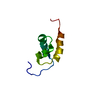
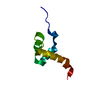
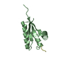
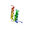
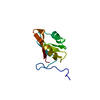
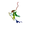
X-ray structure of NdhS from T. elongatus
Components
Tlr0636 protein
Keywords
ELECTRON TRANSPORT / NADH dehydrogenase-like complex / NDH-1 / cyclic electron flow (CEF) / Ferredoxin
Function / homology
NADH dehydrogenase-like complex, subunit S / NAD(P)H dehydrogenase subunit S / photosynthetic electron transport chain / SH3 type barrels. - #140 / SH3 type barrels. / Roll / Mainly Beta / ACETIC ACID / Tlr0636 protein
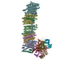
Journal: Science / Year: 2019 Title: Structural adaptations of photosynthetic complex I enable ferredoxin-dependent electron transfer. Authors: Jan M Schuller / James A Birrell / Hideaki Tanaka / Tsuyoshi Konuma / Hannes Wulfhorst / Nicholas Cox / Sandra K Schuller / Jacqueline Thiemann / Wolfgang Lubitz / Pierre Sétif / Takahisa ...Authors: Jan M Schuller / James A Birrell / Hideaki Tanaka / Tsuyoshi Konuma / Hannes Wulfhorst / Nicholas Cox / Sandra K Schuller / Jacqueline Thiemann / Wolfgang Lubitz / Pierre Sétif / Takahisa Ikegami / Benjamin D Engel / Genji Kurisu / Marc M Nowaczyk / Abstract: Photosynthetic complex I enables cyclic electron flow around photosystem I, a regulatory mechanism for photosynthetic energy conversion. We report a 3.3-angstrom-resolution cryo-electron microscopy ...Photosynthetic complex I enables cyclic electron flow around photosystem I, a regulatory mechanism for photosynthetic energy conversion. We report a 3.3-angstrom-resolution cryo-electron microscopy structure of photosynthetic complex I from the cyanobacterium The model reveals structural adaptations that facilitate binding and electron transfer from the photosynthetic electron carrier ferredoxin. By mimicking cyclic electron flow with isolated components in vitro, we demonstrate that ferredoxin directly mediates electron transfer between photosystem I and complex I, instead of using intermediates such as NADPH (the reduced form of nicotinamide adenine dinucleotide phosphate). A large rate constant for association of ferredoxin to complex I indicates efficient recognition, with the protein subunit NdhS being the key component in this process.
Resolution: 1.9→49.71 Å / Cor.coef. Fo:Fc: 0.933 / Cor.coef. Fo:Fc free: 0.931 / SU B: 1.858 / SU ML: 0.057 / Cross valid method: THROUGHOUT / ESU R: 0.025 / ESU R Free: 0.024 / Details: HYDROGENS HAVE BEEN ADDED IN THE RIDING POSITIONS
Rfactor
Num. reflection
% reflection
Selection details
Rfree
0.23783
960
5 %
RANDOM
Rwork
0.21858
-
-
-
obs
0.21957
18235
99.95 %
-
Solvent computation
Ion probe radii: 0.8 Å / Shrinkage radii: 0.8 Å / VDW probe radii: 1.2 Å
 Movie
Movie Controller


 Open data
 Basic information
 Components
 Keywords
 ELECTRON TRANSPORT / NADH dehydrogenase-like complex / NDH-1 / cyclic electron flow (CEF) /
  Function and homology information

 Authors
 Japan, 1items
  Citation


 Structure visualization
 Downloads & links
 Other downloads
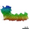


 PDBj
PDBj

 Assembly


 Mass: 60.052 Da / Num. of mol.: 2 / Source method: obtained synthetically / Formula: C2H4O2
 Mass: 18.015 Da / Num. of mol.: 106 / Source method: isolated from a natural source / Formula: H2O
 Sample preparation
 Processing