 Movie
Movie Controller

[English] 日本語
 Yorodumi
- PDB-6a6n: Crystal structure of an inward-open apo state of the eukaryotic A... -
+ Open data

- Basic information
| Entry | Database: PDB / ID: 6a6n | ||||||
|---|---|---|---|---|---|---|---|
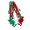
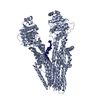
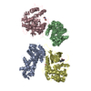
| Title | Crystal structure of an inward-open apo state of the eukaryotic ABC multidrug transporter CmABCB1 | ||||||
 Components Components | ATP-binding cassette, sub-family B, member 1 ATP-binding cassette transporter ATP-binding cassette transporter | ||||||
 Keywords Keywords | TRANSPORT PROTEIN / ALPHA-HELICAL | ||||||
| Function / homology |  Function and homology information Function and homology informationABC-type transporter activity / chloroplast / ATP hydrolysis activity / ATP binding / membraneSimilarity search - Function | ||||||
| Biological species |  Cyanidioschyzon merolae strain 10D (eukaryote) Cyanidioschyzon merolae strain 10D (eukaryote) | ||||||
| Method | X-RAY DIFFRACTION / SYNCHROTRON / MOLECULAR REPLACEMENT / Resolution: 3.02 Å | ||||||
 Authors Authors | Kato, H. / Nakatsu, T. / Kodan, A. | ||||||
 Citation Citation | Journal: Nat Commun / Year: 2019 Title: Inward- and outward-facing X-ray crystal structures of homodimeric P-glycoprotein CmABCB1. Authors: Kodan, A. / Yamaguchi, T. / Nakatsu, T. / Matsuoka, K. / Kimura, Y. / Ueda, K. / Kato, H. | ||||||
| History |
|
- Structure visualization
| Structure viewer | Molecule: MolmilJmol/JSmol |
|---|
- Downloads & links
-Download
| PDBx/mmCIF format | 6a6n.cif.gz | 129.8 KB | Display | PDBx/mmCIF format |
|---|---|---|---|---|
| PDB format | pdb6a6n.ent.gz | 98.1 KB | Display | PDB format |
| PDBx/mmJSON format | 6a6n.json.gz | Tree view | PDBx/mmJSON format | |
| Others |  Other downloads Other downloads |
-Validation report
| Arichive directory | https://data.pdbj.org/pub/pdb/validation_reports/a6/6a6nftp://data.pdbj.org/pub/pdb/validation_reports/a6/6a6n | HTTPS FTP |
|---|
-Related structure data
| Related structure data |  6a6mC 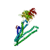 3wmeS S: Starting model for refinement C: citing same article ( |
|---|---|
| Similar structure data |


-Links
 PDBj
PDBj

- Assembly
| Deposited unit | 
| ||||||||
|---|---|---|---|---|---|---|---|---|---|
| 1 | 
| ||||||||
| Unit cell |
|
-Components
| #1: Protein | ATP-binding cassette transporter Mass: 66046.609 Da / Num. of mol.: 1 / Mutation: Q147A, T381A Source method: isolated from a genetically manipulated source Source: (gene. exp.) Cyanidioschyzon merolae strain 10D (eukaryote)Strain: 10D / Gene: CYME_CMD148C / Plasmid: pABC3 / Production host:  Saccharomyces cerevisiae (brewer's yeast) / Strain (production host): AD1-8u- / References: UniProt: M1VAN7 Saccharomyces cerevisiae (brewer's yeast) / Strain (production host): AD1-8u- / References: UniProt: M1VAN7 | ||||
|---|---|---|---|---|---|
| #2: Sugar | ChemComp-DMU /   Type: D-saccharide / Mass: 482.562 Da / Num. of mol.: 4 Type: D-saccharide / Mass: 482.562 Da / Num. of mol.: 4Source method: isolated from a genetically manipulated source Formula: C22H42O11 / Comment: detergent*YM #3: Chemical | ChemComp-TRS / | Tris  Mass: 122.143 Da / Num. of mol.: 1 / Source method: obtained synthetically / Formula: C4H12NO3 / Comment: pH buffer*YM Mass: 122.143 Da / Num. of mol.: 1 / Source method: obtained synthetically / Formula: C4H12NO3 / Comment: pH buffer*YM#4: Water | ChemComp-HOH / | Water Mass: 18.015 Da / Num. of mol.: 13 / Source method: isolated from a natural source / Formula: H2O Mass: 18.015 Da / Num. of mol.: 13 / Source method: isolated from a natural source / Formula: H2O |
-Experimental details
-Experiment
| Experiment | Method: X-RAY DIFFRACTION / Number of used crystals: 1 |
|---|
- Sample preparation
| Crystal | Density Matthews: 3.85 Å3/Da / Density % sol: 68.08 % |
|---|---|
| Crystal grow | Temperature: 293 K / Method: vapor diffusion, sitting drop / Details: 14% PEG 2000 MME, 100mM magnesium nitrate |
-Data collection
| Diffraction | Mean temperature: 100 K |
|---|---|
| Diffraction source | Source: SYNCHROTRON / Site: SPring-8  / Beamline: BL41XU / Wavelength: 1 Å / Beamline: BL41XU / Wavelength: 1 Å |
| Detector | Type: DECTRIS PILATUS3 S 6M / Detector: PIXEL / Date: Dec 19, 2014 |
| Radiation | Monochromator: Si / Protocol: SINGLE WAVELENGTH / Monochromatic (M) / Laue (L): M / Scattering type: x-ray |
| Radiation wavelength | Wavelength: 1 Å / Relative weight: 1 |
| Reflection | Resolution: 3.02→47.14 Å / Num. obs: 19238 / % possible obs: 99.6 % / Redundancy: 9.98 % / Rsym value: 0.102 / Net I/σ(I): 14.3 |
| Reflection shell | Resolution: 3.02→3.1 Å |
- Processing
| Software |
| ||||||||||||||||||||||||||||||||||||||||||||||||||||||||||||||||||||||||||||||||||||||||||||||||||||||||||||||||||||||||||||||||||||||||||||||||||||||||||||||||||||||||||||||||||||||
|---|---|---|---|---|---|---|---|---|---|---|---|---|---|---|---|---|---|---|---|---|---|---|---|---|---|---|---|---|---|---|---|---|---|---|---|---|---|---|---|---|---|---|---|---|---|---|---|---|---|---|---|---|---|---|---|---|---|---|---|---|---|---|---|---|---|---|---|---|---|---|---|---|---|---|---|---|---|---|---|---|---|---|---|---|---|---|---|---|---|---|---|---|---|---|---|---|---|---|---|---|---|---|---|---|---|---|---|---|---|---|---|---|---|---|---|---|---|---|---|---|---|---|---|---|---|---|---|---|---|---|---|---|---|---|---|---|---|---|---|---|---|---|---|---|---|---|---|---|---|---|---|---|---|---|---|---|---|---|---|---|---|---|---|---|---|---|---|---|---|---|---|---|---|---|---|---|---|---|---|---|---|---|---|
| Refinement | Method to determine structure: MOLECULAR REPLACEMENT Starting model: 3WME Resolution: 3.02→47.14 Å / Cor.coef. Fo:Fc: 0.909 / Cor.coef. Fo:Fc free: 0.839 / SU B: 22.948 / SU ML: 0.398 / Cross valid method: THROUGHOUT / ESU R: 7.819 / ESU R Free: 0.422 / Details: HYDROGENS HAVE BEEN USED IF PRESENT IN THE INPUT
| ||||||||||||||||||||||||||||||||||||||||||||||||||||||||||||||||||||||||||||||||||||||||||||||||||||||||||||||||||||||||||||||||||||||||||||||||||||||||||||||||||||||||||||||||||||||
| Solvent computation | Ion probe radii: 0.8 Å / Shrinkage radii: 0.8 Å / VDW probe radii: 1.2 Å | ||||||||||||||||||||||||||||||||||||||||||||||||||||||||||||||||||||||||||||||||||||||||||||||||||||||||||||||||||||||||||||||||||||||||||||||||||||||||||||||||||||||||||||||||||||||
| Displacement parameters | Biso mean: 90.306 Å2
| ||||||||||||||||||||||||||||||||||||||||||||||||||||||||||||||||||||||||||||||||||||||||||||||||||||||||||||||||||||||||||||||||||||||||||||||||||||||||||||||||||||||||||||||||||||||
| Refinement step | Cycle: 1 / Resolution: 3.02→47.14 Å
| ||||||||||||||||||||||||||||||||||||||||||||||||||||||||||||||||||||||||||||||||||||||||||||||||||||||||||||||||||||||||||||||||||||||||||||||||||||||||||||||||||||||||||||||||||||||
| Refine LS restraints |
|