| Entry | Database: PDB / ID: 6a2f
|
|---|
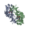
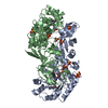
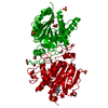
| Title | Crystal structure of biosynthetic alanine racemase from Pseudomonas aeruginosa |
|---|
 Components Components | Alanine racemase, biosynthetic |
|---|
 Keywords Keywords | ISOMERASE / alanine racemase / Alr / Pseudomonas aeruginosa / L-alanine / D-alanine |
|---|
| Function / homology |  Function and homology information Function and homology information
alanine racemase / D-alanine biosynthetic process / alanine racemase activity / peptidoglycan biosynthetic process / cell wall organization / pyridoxal phosphate binding / regulation of cell shape / cytosolSimilarity search - Function Alanine racemase, pyridoxal-phosphate attachment site / Alanine racemase pyridoxal-phosphate attachment site. / Alanine racemase / Alanine racemase, C-terminal / Alanine racemase, C-terminal domain / Alanine racemase, C-terminal domain / Alanine racemase, N-terminal / Alanine racemase, N-terminal domain / Lyase, Ornithine Decarboxylase; Chain A, domain 1 / Lyase, Ornithine Decarboxylase; Chain A, domain 1 ...Alanine racemase, pyridoxal-phosphate attachment site / Alanine racemase pyridoxal-phosphate attachment site. / Alanine racemase / Alanine racemase, C-terminal / Alanine racemase, C-terminal domain / Alanine racemase, C-terminal domain / Alanine racemase, N-terminal / Alanine racemase, N-terminal domain / Lyase, Ornithine Decarboxylase; Chain A, domain 1 / Lyase, Ornithine Decarboxylase; Chain A, domain 1 / Alanine racemase / Alanine racemase/group IV decarboxylase, C-terminal / PLP-binding barrel / TIM Barrel / Alpha-Beta Barrel / Beta Barrel / Mainly Beta / Alpha BetaSimilarity search - Domain/homology |
|---|
| Biological species |  Pseudomonas aeruginosa PAO1 (bacteria) Pseudomonas aeruginosa PAO1 (bacteria) |
|---|
| Method | X-RAY DIFFRACTION / MOLECULAR REPLACEMENT / Resolution: 2.5 Å |
|---|
 Authors Authors | Dong, H. / Li, S. |
|---|
 Citation Citation | Journal: Biochem.Biophys.Res.Commun. / Year: 2018
Title: Enzymatic characterization and crystal structure of biosynthetic alanine racemase from Pseudomonas aeruginosa PAO1.
Authors: Dong, H. / Han, Q. / Guo, Y. / Ju, J. / Wang, S. / Yuan, C. / Long, W. / He, X. / Xu, S. / Li, S. |
|---|
| History | | Deposition | Jun 11, 2018 | Deposition site: PDBJ / Processing site: PDBJ |
|---|
| Revision 1.0 | Aug 8, 2018 | Provider: repository / Type: Initial release |
|---|
| Revision 1.1 | Jan 29, 2020 | Group: Database references / Category: citation / citation_author
Item: _citation.country / _citation.journal_abbrev ..._citation.country / _citation.journal_abbrev / _citation.journal_id_ASTM / _citation.journal_id_CSD / _citation.journal_id_ISSN / _citation.journal_volume / _citation.page_first / _citation.page_last / _citation.pdbx_database_id_DOI / _citation.pdbx_database_id_PubMed / _citation.title / _citation.year |
|---|
| Revision 1.2 | Nov 22, 2023 | Group: Data collection / Database references / Refinement description
Category: chem_comp_atom / chem_comp_bond ...chem_comp_atom / chem_comp_bond / database_2 / pdbx_initial_refinement_model
Item: _database_2.pdbx_DOI / _database_2.pdbx_database_accession |
|---|
|
|---|
 Movie
Movie Controller

 Yorodumi
 Open data
 Basic information
 Structure visualization
 Downloads & links
 Other downloads


 PDBj
PDBj Assembly
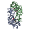

 Mass: 102.046 Da / Num. of mol.: 2 / Source method: obtained synthetically / Formula: C3H2O4
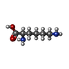
 Type: D-peptide linking / Mass: 146.188 Da / Num. of mol.: 2 / Source method: obtained synthetically / Formula: C6H14N2O2
 Mass: 59.044 Da / Num. of mol.: 2 / Source method: obtained synthetically / Formula: C2H3O2
 Mass: 18.015 Da / Num. of mol.: 344 / Source method: isolated from a natural source / Formula: H2O
 Sample preparation
 Processing