 Movie
Movie Controller

[English] 日本語
 Yorodumi
- PDB-5zyg: Crystal structure of CERT START domain in complex with compound B5 -
+ Open data

- Basic information
| Entry | Database: PDB / ID: 5zyg | ||||||
|---|---|---|---|---|---|---|---|
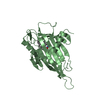
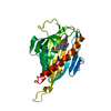
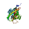
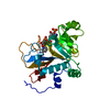
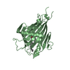
| Title | Crystal structure of CERT START domain in complex with compound B5 | ||||||
 Components Components | LIPID-TRANSFER PROTEIN CERT | ||||||
 Keywords Keywords | LIPID TRANSPORT / CERT / PH /  START / COMPLEX START / COMPLEX | ||||||
| Function / homology |  Function and homology information Function and homology informationintermembrane sphingolipid transfer / ceramide transfer activity / ceramide transport / : / ceramide 1-phosphate binding / ceramide 1-phosphate transfer activity / ER to Golgi ceramide transport / ceramide binding / Sphingolipid de novo biosynthesis / intermembrane lipid transfer ...intermembrane sphingolipid transfer / ceramide transfer activity / ceramide transport / : / ceramide 1-phosphate binding / ceramide 1-phosphate transfer activity / ER to Golgi ceramide transport / ceramide binding / Sphingolipid de novo biosynthesis / intermembrane lipid transfer / ceramide metabolic process / endoplasmic reticulum organization / phosphatidylinositol-4-phosphate binding / lipid homeostasis / heart morphogenesis / response to endoplasmic reticulum stress / muscle contraction / cell morphogenesis / kinase activity / in utero embryonic development / cell population proliferation / immune response / endoplasmic reticulum membrane / Golgi apparatus / signal transduction / mitochondrion / nucleoplasm / identical protein binding / cytosolSimilarity search - Function | ||||||
| Biological species |  Homo sapiens (human) Homo sapiens (human) | ||||||
| Method | X-RAY DIFFRACTION / Resolution: 1.8 Å | ||||||
 Authors Authors | Suzuki, M. / Nakao, N. / Ueno, M. / Sakai, S. / Egawa, D. / Hanzawa, H. / Kawasaki, S. / Kumagai, K. / Kobayashi, S. / Hanada, K. | ||||||
 Citation Citation | Journal: Commun Chem / Year: 2019 Title: Natural ligand-nonmimetic inhibitors of the lipid-transfer protein CERT Authors: Nakao, N. / Ueno, M. / Sakai, S. / Egawa, D. / Hanzawa, H. / Kawasaki, S. / Kumagai, K. / Suzuki, M. / Kobayashi, S. / Hanada, K. | ||||||
| History |
|
- Structure visualization
| Structure viewer | Molecule: MolmilJmol/JSmol |
|---|
- Downloads & links
-Download
| PDBx/mmCIF format | 5zyg.cif.gz | 68.3 KB | Display | PDBx/mmCIF format |
|---|---|---|---|---|
| PDB format | pdb5zyg.ent.gz | 48.7 KB | Display | PDB format |
| PDBx/mmJSON format | 5zyg.json.gz | Tree view | PDBx/mmJSON format | |
| Others |  Other downloads Other downloads |
-Validation report
| Arichive directory | https://data.pdbj.org/pub/pdb/validation_reports/zy/5zygftp://data.pdbj.org/pub/pdb/validation_reports/zy/5zyg | HTTPS FTP |
|---|
-Related structure data
| Related structure data |  5zyhC  5zyiC  5zyjC  5zykC  5zylC  5zymC 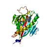 6iezC  6if0C  6j0oC  6j81C C: citing same article ( |
|---|---|
| Similar structure data |

-Links
 PDBj
PDBj
- Assembly
| Deposited unit | 
| ||||||||
|---|---|---|---|---|---|---|---|---|---|
| 1 |
| ||||||||
| Unit cell |
|
-Components
| #1: Protein | Mass: 27056.781 Da / Num. of mol.: 1 / Fragment: UNP residues 364-598 Source method: isolated from a genetically manipulated source Source: (gene. exp.) Homo sapiens (human) / Gene: CERT / Plasmid: pET28b-His-3C-CERT START / Production host:  Escherichia coli BL21(DE3) (bacteria) / Strain (production host): BL21(DE3) / References: UniProt: Q9Y5P4 Escherichia coli BL21(DE3) (bacteria) / Strain (production host): BL21(DE3) / References: UniProt: Q9Y5P4 |
|---|---|
| #2: Chemical | ChemComp-9N3 /   Mass: 423.568 Da / Num. of mol.: 1 / Source method: obtained synthetically / Formula: C25H29NO3S Mass: 423.568 Da / Num. of mol.: 1 / Source method: obtained synthetically / Formula: C25H29NO3S |
| #3: Water | ChemComp-HOH / Water Mass: 18.015 Da / Num. of mol.: 218 / Source method: isolated from a natural source / Formula: H2O Mass: 18.015 Da / Num. of mol.: 218 / Source method: isolated from a natural source / Formula: H2O |
| Sequence details | Two residues GLY362A and PRO363A are originated from protease site after N-terminal affinity tag. ...Two residues GLY362A and PRO363A are originated from protease site after N-terminal affinity tag. THIS SEQUENCE CORRESPOND |
-Experimental details
-Experiment
| Experiment | Method: X-RAY DIFFRACTION / Number of used crystals: 1 |
|---|
- Sample preparation
| Crystal | Density Matthews: 2.55 Å3/Da / Density % sol: 51.75 % |
|---|---|
| Crystal grow | Temperature: 293 K / Method: vapor diffusion, sitting drop / pH: 5.9 Details: 0.1M trisodium citrate/HCl buffer, pH 5.9, 24% PEG3350 and 0.2% n-octyl-beta-D-glucoside |
-Data collection
| Diffraction | Mean temperature: 100 K | ||||||||||||||||||||||||
|---|---|---|---|---|---|---|---|---|---|---|---|---|---|---|---|---|---|---|---|---|---|---|---|---|---|
| Diffraction source | Source: ROTATING ANODE / Type: RIGAKU FR-E SUPERBRIGHT / Wavelength: 1.5418 Å | ||||||||||||||||||||||||
| Detector | Type: RIGAKU RAXIS IV / Detector: IMAGE PLATE / Date: Jan 7, 2016 | ||||||||||||||||||||||||
| Radiation | Protocol: SINGLE WAVELENGTH / Monochromatic (M) / Laue (L): M / Scattering type: x-ray | ||||||||||||||||||||||||
| Radiation wavelength | Wavelength: 1.5418 Å / Relative weight: 1 | ||||||||||||||||||||||||
| Reflection | Resolution: 1.8→39.01 Å / Num. obs: 26757 / % possible obs: 99.7 % / Redundancy: 3.4 % / CC1/2: 0.997 / Rmerge(I) obs: 0.053 / Rpim(I) all: 0.034 / Rrim(I) all: 0.063 / Net I/σ(I): 12.6 / Num. measured all: 90169 / Scaling rejects: 39 | ||||||||||||||||||||||||
| Reflection shell | Diffraction-ID: 1
|
- Processing
| Software |
| |||||||||||||||||||||||||||||||||||||||||||||
|---|---|---|---|---|---|---|---|---|---|---|---|---|---|---|---|---|---|---|---|---|---|---|---|---|---|---|---|---|---|---|---|---|---|---|---|---|---|---|---|---|---|---|---|---|---|---|
| Refinement | Resolution: 1.8→25 Å / Cor.coef. Fo:Fc: 0.954 / Cor.coef. Fo:Fc free: 0.94 / SU B: 2.726 / SU ML: 0.083 / Cross valid method: THROUGHOUT / σ(F): 0 / ESU R: 0.127 / ESU R Free: 0.121 / Details: U VALUES : REFINED INDIVIDUALLY
| |||||||||||||||||||||||||||||||||||||||||||||
| Solvent computation | Ion probe radii: 0.8 Å / Shrinkage radii: 0.8 Å / VDW probe radii: 1.2 Å | |||||||||||||||||||||||||||||||||||||||||||||
| Displacement parameters | Biso max: 79.73 Å2 / Biso mean: 25.066 Å2 / Biso min: 11.25 Å2
| |||||||||||||||||||||||||||||||||||||||||||||
| Refinement step | Cycle: final / Resolution: 1.8→25 Å
| |||||||||||||||||||||||||||||||||||||||||||||
| Refine LS restraints |
| |||||||||||||||||||||||||||||||||||||||||||||
| LS refinement shell | Resolution: 1.8→1.863 Å / Rfactor Rfree error: 0 / Total num. of bins used: 15
|
