 Movie
Movie Controller


+ Open data

- Basic information
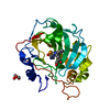
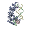
| Entry | Database: PDB / ID: 5zof | ||||||
|---|---|---|---|---|---|---|---|
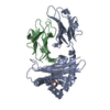
| Title | Crystal Structure of D181A/R192F hFen1 in complex with DNA | ||||||
 Components Components |
| ||||||
 Keywords Keywords | HYDROLASE/DNA / flap endonuclease: gap endonuclease: methylation: posttranslational modification /  DNA BINDING PROTEIN / HYDROLASE-DNA complex DNA BINDING PROTEIN / HYDROLASE-DNA complex | ||||||
| Function / homology |  Function and homology information Function and homology informationpositive regulation of sister chromatid cohesion / flap endonuclease activity / telomere maintenance via semi-conservative replication / double-stranded DNA exodeoxyribonuclease activity / nucleic acid metabolic process / 5'-flap endonuclease activity / DNA replication, removal of RNA primer / 5'-3' exonuclease activity / UV protection / Removal of the Flap Intermediate ...positive regulation of sister chromatid cohesion / flap endonuclease activity / telomere maintenance via semi-conservative replication / double-stranded DNA exodeoxyribonuclease activity / nucleic acid metabolic process / 5'-flap endonuclease activity / DNA replication, removal of RNA primer / 5'-3' exonuclease activity / UV protection / Removal of the Flap Intermediate / HDR through MMEJ (alt-NHEJ) / Removal of the Flap Intermediate from the C-strand / exonuclease activity / Early Phase of HIV Life Cycle / PCNA-Dependent Long Patch Base Excision Repair / POLB-Dependent Long Patch Base Excision Repair / base-excision repair, gap-filling / double-strand break repair via homologous recombination / memory / double-strand break repair / RNA-DNA hybrid ribonuclease activity / manganese ion binding / double-stranded DNA binding / endonuclease activity / DNA replication / damaged DNA binding / chromosome, telomeric region / Hydrolases; Acting on ester bonds / DNA repair / nucleolus / magnesium ion binding / protein-containing complex / mitochondrion / DNA binding / nucleoplasm / membrane / nucleusSimilarity search - Function | ||||||
| Biological species |  Homo sapiens (human) Homo sapiens (human)synthetic construct (others) | ||||||
| Method | X-RAY DIFFRACTION / SYNCHROTRON / MOLECULAR REPLACEMENT / Resolution: 2.249 Å | ||||||
 Authors Authors | Han, W. / Hua, Y. / Zhao, Y. | ||||||
| Funding support |  China, 1items China, 1items
| ||||||
 Citation Citation | Journal: Nucleic Acids Res. / Year: 2018 Title: Structural basis of 5' flap recognition and protein-protein interactions of human flap endonuclease 1. Authors: Xu, H. / Shi, R. / Han, W. / Cheng, J. / Xu, X. / Cheng, K. / Wang, L. / Tian, B. / Zheng, L. / Shen, B. / Hua, Y. / Zhao, Y. | ||||||
| History |
|
- Structure visualization
| Structure viewer | Molecule: MolmilJmol/JSmol |
|---|
- Downloads & links
-Download
| PDBx/mmCIF format | 5zof.cif.gz | 96.5 KB | Display | PDBx/mmCIF format |
|---|---|---|---|---|
| PDB format | pdb5zof.ent.gz | 67.7 KB | Display | PDB format |
| PDBx/mmJSON format | 5zof.json.gz | Tree view | PDBx/mmJSON format | |
| Others |  Other downloads Other downloads |
-Validation report
| Arichive directory | https://data.pdbj.org/pub/pdb/validation_reports/zo/5zofftp://data.pdbj.org/pub/pdb/validation_reports/zo/5zof | HTTPS FTP |
|---|
-Related structure data
| Related structure data | 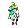 5zodC  5zoeC  5zogC  3q8kS S: Starting model for refinement C: citing same article ( |
|---|---|
| Similar structure data |


-Links
 PDBj
PDBj


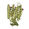
- Assembly
| Deposited unit | 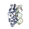
| ||||||||
|---|---|---|---|---|---|---|---|---|---|
| 1 |
| ||||||||
| Unit cell |
|
-Components
-Protein , 1 types, 1 molecules A
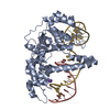
| #1: Protein | Flap structure-specific endonuclease 1 / FEN-1 / DNase IV / Flap structure-specific endonuclease 1 / Maturation factor 1 / hFEN-1 Mass: 37613.262 Da / Num. of mol.: 1 / Fragment: nuclease core (1-333) / Mutation: D181A, R192F Source method: isolated from a genetically manipulated source Source: (gene. exp.) Homo sapiens (human) / Gene: FEN1, RAD2 / Plasmid: pET28b / Production host:  Escherichia coli (E. coli) / Strain (production host): BL21(DE3) PlysS Escherichia coli (E. coli) / Strain (production host): BL21(DE3) PlysSReferences: UniProt: P39748, Hydrolases; Acting on ester bonds |
|---|
-DNA chain , 3 types, 3 molecules BCD
| #2: DNA chain | Mass: 5477.543 Da / Num. of mol.: 1 / Source method: obtained synthetically / Source: (synth.) synthetic construct (others) |
|---|---|
| #3: DNA chain | Mass: 2363.556 Da / Num. of mol.: 1 / Source method: obtained synthetically / Source: (synth.) synthetic construct (others) |
| #4: DNA chain | Mass: 4344.840 Da / Num. of mol.: 1 / Source method: obtained synthetically / Source: (synth.) synthetic construct (others) |
-Non-polymers , 2 types, 11 molecules 

| #5: Chemical | ChemComp-K /  Mass: 39.098 Da / Num. of mol.: 1 / Source method: obtained synthetically / Formula: K Mass: 39.098 Da / Num. of mol.: 1 / Source method: obtained synthetically / Formula: K |
|---|---|
| #6: Water | ChemComp-HOH / WaterMass: 18.015 Da / Num. of mol.: 10 / Source method: isolated from a natural source / Formula: H2O |
-Experimental details
-Experiment
| Experiment | Method: X-RAY DIFFRACTION / Number of used crystals: 1 |
|---|
- Sample preparation
| Crystal | Density Matthews: 2.28 Å3/Da / Density % sol: 46.01 % |
|---|---|
| Crystal grow | Temperature: 298 K / Method: vapor diffusion, sitting drop / pH: 7.8 / Details: PEG 3350, MgCl2, Tris, KCl |
-Data collection
| Diffraction | Mean temperature: 95 K | ||||||||||||||||||||||||||||||||||||||||||||||||||||||||||||||||||||||||||||||||||||||||||||||||||||||||||||||||||||||||||||||||||||||||||||||||||||||||||||||||||||||||
|---|---|---|---|---|---|---|---|---|---|---|---|---|---|---|---|---|---|---|---|---|---|---|---|---|---|---|---|---|---|---|---|---|---|---|---|---|---|---|---|---|---|---|---|---|---|---|---|---|---|---|---|---|---|---|---|---|---|---|---|---|---|---|---|---|---|---|---|---|---|---|---|---|---|---|---|---|---|---|---|---|---|---|---|---|---|---|---|---|---|---|---|---|---|---|---|---|---|---|---|---|---|---|---|---|---|---|---|---|---|---|---|---|---|---|---|---|---|---|---|---|---|---|---|---|---|---|---|---|---|---|---|---|---|---|---|---|---|---|---|---|---|---|---|---|---|---|---|---|---|---|---|---|---|---|---|---|---|---|---|---|---|---|---|---|---|---|---|---|---|
| Diffraction source | Source: SYNCHROTRON / Site: SSRF / Beamline: BL17U1 / Wavelength: 1 Å | ||||||||||||||||||||||||||||||||||||||||||||||||||||||||||||||||||||||||||||||||||||||||||||||||||||||||||||||||||||||||||||||||||||||||||||||||||||||||||||||||||||||||
| Detector | Type: ADSC QUANTUM 315r / Detector: CCD / Date: Dec 4, 2016 | ||||||||||||||||||||||||||||||||||||||||||||||||||||||||||||||||||||||||||||||||||||||||||||||||||||||||||||||||||||||||||||||||||||||||||||||||||||||||||||||||||||||||
| Radiation | Protocol: SINGLE WAVELENGTH / Monochromatic (M) / Laue (L): M / Scattering type: x-ray | ||||||||||||||||||||||||||||||||||||||||||||||||||||||||||||||||||||||||||||||||||||||||||||||||||||||||||||||||||||||||||||||||||||||||||||||||||||||||||||||||||||||||
| Radiation wavelength | Wavelength: 1 Å / Relative weight: 1 | ||||||||||||||||||||||||||||||||||||||||||||||||||||||||||||||||||||||||||||||||||||||||||||||||||||||||||||||||||||||||||||||||||||||||||||||||||||||||||||||||||||||||
| Reflection | Resolution: 2.24→27.82 Å / Num. obs: 22092 / % possible obs: 98.8 % / Redundancy: 5.74 % / Biso Wilson estimate: 46.8 Å2 / CC1/2: 0.994 / Rmerge(I) obs: 0.091 / Rrim(I) all: 0.101 / Χ2: 0.972 / Net I/σ(I): 11.12 | ||||||||||||||||||||||||||||||||||||||||||||||||||||||||||||||||||||||||||||||||||||||||||||||||||||||||||||||||||||||||||||||||||||||||||||||||||||||||||||||||||||||||
| Reflection shell | Diffraction-ID: 1
|
- Processing
| Software |
| |||||||||||||||||||||||||||||||||||||||||||||||||||||||||||||||
|---|---|---|---|---|---|---|---|---|---|---|---|---|---|---|---|---|---|---|---|---|---|---|---|---|---|---|---|---|---|---|---|---|---|---|---|---|---|---|---|---|---|---|---|---|---|---|---|---|---|---|---|---|---|---|---|---|---|---|---|---|---|---|---|---|
| Refinement | Method to determine structure: MOLECULAR REPLACEMENT Starting model: 3Q8K Resolution: 2.249→27.82 Å / SU ML: 0.35 / Cross valid method: THROUGHOUT / σ(F): 1.36 / Phase error: 32.29 / Stereochemistry target values: ML
| |||||||||||||||||||||||||||||||||||||||||||||||||||||||||||||||
| Solvent computation | Shrinkage radii: 0.9 Å / VDW probe radii: 1.11 Å / Solvent model: FLAT BULK SOLVENT MODEL | |||||||||||||||||||||||||||||||||||||||||||||||||||||||||||||||
| Displacement parameters | Biso max: 133.83 Å2 / Biso mean: 56.5319 Å2 / Biso min: 24.57 Å2 | |||||||||||||||||||||||||||||||||||||||||||||||||||||||||||||||
| Refinement step | Cycle: final / Resolution: 2.249→27.82 Å
| |||||||||||||||||||||||||||||||||||||||||||||||||||||||||||||||
| LS refinement shell | Refine-ID: X-RAY DIFFRACTION / Rfactor Rfree error: 0 / Total num. of bins used: 8
|
