 Movie
Movie Controller


+ Open data

- Basic information
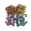
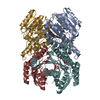
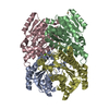
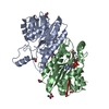
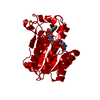
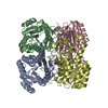
| Entry | Database: PDB / ID: 5zfm | |||||||||
|---|---|---|---|---|---|---|---|---|---|---|
| Title | Ketoreductase LbCR mutant - M6 | |||||||||
 Components Components | 3-oxoacyl-acyl carrier protein reductase | |||||||||
 Keywords Keywords |  OXIDOREDUCTASE / ketoreductase / short-chain dehydrogenase/reductase (SDR) OXIDOREDUCTASE / ketoreductase / short-chain dehydrogenase/reductase (SDR) | |||||||||
| Function / homology |  Function and homology information Function and homology information | |||||||||
| Biological species |  Lactobacillus brevis (bacteria) Lactobacillus brevis (bacteria) | |||||||||
| Method | X-RAY DIFFRACTION / SYNCHROTRON / MOLECULAR REPLACEMENT / Resolution: 2 Å | |||||||||
 Authors Authors | Gong, X.M. / Zheng, G.W. / Xu, J.H. | |||||||||
| Funding support |  China, 2items China, 2items
| |||||||||
 Citation Citation | Journal: Acs Catalysis / Year: 2019 Title: Development of an Engineered Ketoreductase with Simultaneously Improved Thermostability and Activity for Making a Bulky Atorvastatin Precursor Authors: Gong, X.M. / Zhen, Q. / Li, F.L. / Zeng, B.B. / Zheng, G.W. / Xu, J.H. | |||||||||
| History |
|
- Structure visualization
| Structure viewer | Molecule: MolmilJmol/JSmol |
|---|
- Downloads & links
-Download
| PDBx/mmCIF format | 5zfm.cif.gz | 220.1 KB | Display | PDBx/mmCIF format |
|---|---|---|---|---|
| PDB format | pdb5zfm.ent.gz | 172.7 KB | Display | PDB format |
| PDBx/mmJSON format | 5zfm.json.gz | Tree view | PDBx/mmJSON format | |
| Others |  Other downloads Other downloads |
-Validation report
| Arichive directory | https://data.pdbj.org/pub/pdb/validation_reports/zf/5zfmftp://data.pdbj.org/pub/pdb/validation_reports/zf/5zfm | HTTPS FTP |
|---|
-Related structure data


-Links
 PDBj
PDBj


- Assembly
| Deposited unit | 
| ||||||||
|---|---|---|---|---|---|---|---|---|---|
| 1 |
| ||||||||
| Unit cell |
|
-Components
| #1: Protein | Mass: 29553.455 Da / Num. of mol.: 4 / Mutation: M154A,A155D,A201D,A202L Source method: isolated from a genetically manipulated source Source: (gene. exp.) Lactobacillus brevis (strain ATCC 367 / JCM 1170) (bacteria)Strain: ATCC 367 / JCM 1170 / Gene: LVIS_0330 Production host: Escherichia coli 'BL21-Gold(DE3)pLysS AG' (bacteria)References: UniProt: Q03TH6 #2: Water | ChemComp-HOH / | Water Mass: 18.015 Da / Num. of mol.: 1184 / Source method: isolated from a natural source / Formula: H2O Mass: 18.015 Da / Num. of mol.: 1184 / Source method: isolated from a natural source / Formula: H2O |
|---|
-Experimental details
-Experiment
| Experiment | Method: X-RAY DIFFRACTION / Number of used crystals: 1 |
|---|
- Sample preparation
| Crystal | Density Matthews: 2.33 Å3/Da / Density % sol: 47.25 % |
|---|---|
| Crystal grow | Temperature: 291.15 K / Method: vapor diffusion, sitting drop Details: PEG 4000, MES buffer, sodium chloride, glycyl-glycyl-glycine |
-Data collection
| Diffraction | Mean temperature: 100 K |
|---|---|
| Diffraction source | Source: SYNCHROTRON / Site: SSRF / Beamline: BL19U1 / Wavelength: 0.97775 Å |
| Detector | Type: DECTRIS PILATUS3 S 6M / Detector: PIXEL / Date: Jun 9, 2017 |
| Radiation | Protocol: SINGLE WAVELENGTH / Monochromatic (M) / Laue (L): M / Scattering type: x-ray |
| Radiation wavelength | Wavelength: 0.97775 Å / Relative weight: 1 |
| Reflection | Resolution: 2→44.28 Å / Num. obs: 72544 / % possible obs: 99.08 % / Redundancy: 6.6 % / Rmerge(I) obs: 0.197 / Net I/σ(I): 10.143 |
| Reflection shell | Resolution: 2→2.071 Å / Rmerge(I) obs: 0.516 |
- Processing
| Software |
| |||||||||||||||||||||||||||||||||||||||||||||||||||||||||||||||||||||||||||||||||||||||||||||||||||||||||||||||||||||||||||||||||||||||||||||||||||||||||||||||||||||||||||||||||||||||||||||
|---|---|---|---|---|---|---|---|---|---|---|---|---|---|---|---|---|---|---|---|---|---|---|---|---|---|---|---|---|---|---|---|---|---|---|---|---|---|---|---|---|---|---|---|---|---|---|---|---|---|---|---|---|---|---|---|---|---|---|---|---|---|---|---|---|---|---|---|---|---|---|---|---|---|---|---|---|---|---|---|---|---|---|---|---|---|---|---|---|---|---|---|---|---|---|---|---|---|---|---|---|---|---|---|---|---|---|---|---|---|---|---|---|---|---|---|---|---|---|---|---|---|---|---|---|---|---|---|---|---|---|---|---|---|---|---|---|---|---|---|---|---|---|---|---|---|---|---|---|---|---|---|---|---|---|---|---|---|---|---|---|---|---|---|---|---|---|---|---|---|---|---|---|---|---|---|---|---|---|---|---|---|---|---|---|---|---|---|---|---|---|
| Refinement | Method to determine structure: MOLECULAR REPLACEMENT / Resolution: 2→44.278 Å / SU ML: 0.2 / Cross valid method: FREE R-VALUE / σ(F): 1.36 / Phase error: 19.62
| |||||||||||||||||||||||||||||||||||||||||||||||||||||||||||||||||||||||||||||||||||||||||||||||||||||||||||||||||||||||||||||||||||||||||||||||||||||||||||||||||||||||||||||||||||||||||||||
| Solvent computation | Shrinkage radii: 0.9 Å / VDW probe radii: 1.11 Å | |||||||||||||||||||||||||||||||||||||||||||||||||||||||||||||||||||||||||||||||||||||||||||||||||||||||||||||||||||||||||||||||||||||||||||||||||||||||||||||||||||||||||||||||||||||||||||||
| Refinement step | Cycle: LAST / Resolution: 2→44.278 Å
| |||||||||||||||||||||||||||||||||||||||||||||||||||||||||||||||||||||||||||||||||||||||||||||||||||||||||||||||||||||||||||||||||||||||||||||||||||||||||||||||||||||||||||||||||||||||||||||
| Refine LS restraints |
| |||||||||||||||||||||||||||||||||||||||||||||||||||||||||||||||||||||||||||||||||||||||||||||||||||||||||||||||||||||||||||||||||||||||||||||||||||||||||||||||||||||||||||||||||||||||||||||
| LS refinement shell |
|
