 Movie
Movie Controller

[English] 日本語
 Yorodumi
- PDB-5zan: Crystal Structure of Aurora-A in complex with a new Quinazoline i... -
+ Open data

- Basic information
| Entry | Database: PDB / ID: 5zan | ||||||
|---|---|---|---|---|---|---|---|
| Title | Crystal Structure of Aurora-A in complex with a new Quinazoline inhibitor | ||||||
 Components Components | Aurora kinase A | ||||||
 Keywords Keywords | TRANSFERASE/INHIBITOR / Drug Design / TRANSFERASE / TRANSFERASE-INHIBITOR complex | ||||||
| Function / homology |  Function and homology information Function and homology informationInteraction between PHLDA1 and AURKA / regulation of centrosome cycle / axon hillock / spindle assembly involved in female meiosis I / cilium disassembly / spindle pole centrosome / positive regulation of oocyte maturation / histone H3S10 kinase activity / chromosome passenger complex / pronucleus ...Interaction between PHLDA1 and AURKA / regulation of centrosome cycle / axon hillock / spindle assembly involved in female meiosis I / cilium disassembly / spindle pole centrosome / positive regulation of oocyte maturation / histone H3S10 kinase activity / chromosome passenger complex / pronucleus / meiotic spindle / mitotic centrosome separation / germinal vesicle / protein localization to centrosome / anterior/posterior axis specification / centrosome localization / neuron projection extension / positive regulation of mitochondrial fission / spindle organization / mitotic spindle pole / SUMOylation of DNA replication proteins / spindle midzone / regulation of G2/M transition of mitotic cell cycle / centriole / protein serine/threonine/tyrosine kinase activity / positive regulation of mitotic cell cycle / AURKA Activation by TPX2 / TP53 Regulates Transcription of Genes Involved in G2 Cell Cycle Arrest / positive regulation of mitotic nuclear division / mitotic spindle organization / ciliary basal body / negative regulation of protein binding / regulation of signal transduction by p53 class mediator / regulation of cytokinesis / molecular function activator activity / liver regeneration / FBXL7 down-regulates AURKA during mitotic entry and in early mitosis / APC/C:Cdh1 mediated degradation of Cdc20 and other APC/C:Cdh1 targeted proteins in late mitosis/early G1 / regulation of protein stability / spindle / mitotic spindle / kinetochore / response to wounding / microtubule cytoskeleton / G2/M transition of mitotic cell cycle / Regulation of PLK1 Activity at G2/M Transition / positive regulation of proteasomal ubiquitin-dependent protein catabolic process / mitotic cell cycle / midbody / proteasome-mediated ubiquitin-dependent protein catabolic process / basolateral plasma membrane / peptidyl-serine phosphorylation / Regulation of TP53 Activity through Phosphorylation / microtubule / postsynaptic density / protein autophosphorylation / non-specific serine/threonine protein kinase / protein kinase activity / protein heterodimerization activity / cell division / negative regulation of gene expression / protein phosphorylation / protein serine kinase activity / centrosome / protein serine/threonine kinase activity / glutamatergic synapse / apoptotic process / ubiquitin protein ligase binding / negative regulation of apoptotic process / protein kinase binding / perinuclear region of cytoplasm / nucleoplasm / ATP binding / nucleus / cytosolSimilarity search - Function | ||||||
| Biological species |  Homo sapiens (human) Homo sapiens (human) | ||||||
| Method | X-RAY DIFFRACTION / SYNCHROTRON / MOLECULAR REPLACEMENT / Resolution: 2.85 Å | ||||||
 Authors Authors | Cheng, B. / Lu, G. / Zhou, H. | ||||||
| Funding support |  China, 1items China, 1items
| ||||||
 Citation Citation | Journal: To Be Published Title: Optimization of Novel Quinazolines as Potent and Orally Bioavailable Aurora Kinase Inhibitors Authors: Long, L. / Peng, W. / Pan, Y. / Cheng, B. / Tu, Z. / Long, Z. / Zhou, H. / Liu, Q. / Lu, G. | ||||||
| History |
|
- Structure visualization
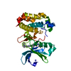
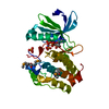
| Structure viewer | Molecule: MolmilJmol/JSmol |
|---|
- Downloads & links
-Download
| PDBx/mmCIF format | 5zan.cif.gz | 119.1 KB | Display | PDBx/mmCIF format |
|---|---|---|---|---|
| PDB format | pdb5zan.ent.gz | 91.1 KB | Display | PDB format |
| PDBx/mmJSON format | 5zan.json.gz | Tree view | PDBx/mmJSON format | |
| Others |  Other downloads Other downloads |
-Validation report
| Arichive directory | https://data.pdbj.org/pub/pdb/validation_reports/za/5zanftp://data.pdbj.org/pub/pdb/validation_reports/za/5zan | HTTPS FTP |
|---|
-Related structure data
| Related structure data |  4utd S: Starting model for refinement |
|---|---|
| Similar structure data |


-Links
 PDBj
PDBj


- Assembly
| Deposited unit | 
| ||||||||
|---|---|---|---|---|---|---|---|---|---|
| 1 |
| ||||||||
| Unit cell |
|
-Components
| #1: Protein | / Aurora 2 / Aurora/IPL1-related kinase 1 / hARK1 / Breast tumor-amplified kinase / Serine/threonine- ...Aurora 2 / Aurora/IPL1-related kinase 1 / hARK1 / Breast tumor-amplified kinase / Serine/threonine-protein kinase 15 / Serine/threonine-protein kinase 6 / Serine/threonine-protein kinase aurora-A Mass: 32560.346 Da / Num. of mol.: 1 / Fragment: Catalytic domain, UNP residues 123-403 Source method: isolated from a genetically manipulated source Source: (gene. exp.) Homo sapiens (human)Gene: AURKA, AIK, AIRK1, ARK1, AURA, AYK1, BTAK, IAK1, STK15, STK6 Production host:  Escherichia coli (E. coli) Escherichia coli (E. coli)References: UniProt: O14965, non-specific serine/threonine protein kinase |
|---|---|
| #2: Chemical | ChemComp-9A6 /   Mass: 425.529 Da / Num. of mol.: 1 / Source method: obtained synthetically / Formula: C25H27N7 / Feature type: SUBJECT OF INVESTIGATION Mass: 425.529 Da / Num. of mol.: 1 / Source method: obtained synthetically / Formula: C25H27N7 / Feature type: SUBJECT OF INVESTIGATION |
-Experimental details
-Experiment
| Experiment | Method: X-RAY DIFFRACTION / Number of used crystals: 1 |
|---|
- Sample preparation
| Crystal | Density Matthews: 2.81 Å3/Da / Density % sol: 56.3 % |
|---|---|
| Crystal grow | Temperature: 293 K / Method: evaporation Details: 0.22M Sodium citrate tribasic dihydrate pH 6.0, 10%(v/v) 2-Propanol, 22(w/v) Polyethylene glycol 4,000 |
-Data collection
| Diffraction | Mean temperature: 100 K |
|---|---|
| Diffraction source | Source: SYNCHROTRON / Site: SSRF / Beamline: BL17U1 / Wavelength: 0.979 Å |
| Detector | Type: ADSC QUANTUM 315 / Detector: CCD / Date: Apr 2, 2016 |
| Radiation | Protocol: SINGLE WAVELENGTH / Monochromatic (M) / Laue (L): M / Scattering type: x-ray |
| Radiation wavelength | Wavelength: 0.979 Å / Relative weight: 1 |
| Reflection | Resolution: 2.85→50 Å / Num. obs: 9017 / % possible obs: 98.8 % / Redundancy: 11.2 % / Rmerge(I) obs: 0.066 / Net I/σ(I): 54.4 |
| Reflection shell | Resolution: 2.85→2.9 Å / Rmerge(I) obs: 0.561 / Mean I/σ(I) obs: 5.8 / Num. unique obs: 428 |
- Processing
| Software |
| ||||||||||||||||||||||||||||||||||||||||||||||||||||||||||||||||||||||||||||||||||||||||||||||||||||||||||||||||||||||||||||||||||||||||||||||||||||||||||||||||||||||||||||||||||||||
|---|---|---|---|---|---|---|---|---|---|---|---|---|---|---|---|---|---|---|---|---|---|---|---|---|---|---|---|---|---|---|---|---|---|---|---|---|---|---|---|---|---|---|---|---|---|---|---|---|---|---|---|---|---|---|---|---|---|---|---|---|---|---|---|---|---|---|---|---|---|---|---|---|---|---|---|---|---|---|---|---|---|---|---|---|---|---|---|---|---|---|---|---|---|---|---|---|---|---|---|---|---|---|---|---|---|---|---|---|---|---|---|---|---|---|---|---|---|---|---|---|---|---|---|---|---|---|---|---|---|---|---|---|---|---|---|---|---|---|---|---|---|---|---|---|---|---|---|---|---|---|---|---|---|---|---|---|---|---|---|---|---|---|---|---|---|---|---|---|---|---|---|---|---|---|---|---|---|---|---|---|---|---|---|
| Refinement | Method to determine structure: MOLECULAR REPLACEMENT Starting model: 4UTD 4utd Resolution: 2.85→50 Å / Cor.coef. Fo:Fc: 0.926 / Cor.coef. Fo:Fc free: 0.909 / SU B: 44.874 / SU ML: 0.388 / Cross valid method: THROUGHOUT / ESU R: 1.494 / ESU R Free: 0.397 / Stereochemistry target values: MAXIMUM LIKELIHOOD / Details: HYDROGENS HAVE BEEN ADDED IN THE RIDING POSITIONS
| ||||||||||||||||||||||||||||||||||||||||||||||||||||||||||||||||||||||||||||||||||||||||||||||||||||||||||||||||||||||||||||||||||||||||||||||||||||||||||||||||||||||||||||||||||||||
| Solvent computation | Ion probe radii: 0.8 Å / Shrinkage radii: 0.8 Å / VDW probe radii: 1.2 Å / Solvent model: MASK | ||||||||||||||||||||||||||||||||||||||||||||||||||||||||||||||||||||||||||||||||||||||||||||||||||||||||||||||||||||||||||||||||||||||||||||||||||||||||||||||||||||||||||||||||||||||
| Displacement parameters | Biso mean: 100.013 Å2
| ||||||||||||||||||||||||||||||||||||||||||||||||||||||||||||||||||||||||||||||||||||||||||||||||||||||||||||||||||||||||||||||||||||||||||||||||||||||||||||||||||||||||||||||||||||||
| Refinement step | Cycle: 1 / Resolution: 2.85→50 Å
| ||||||||||||||||||||||||||||||||||||||||||||||||||||||||||||||||||||||||||||||||||||||||||||||||||||||||||||||||||||||||||||||||||||||||||||||||||||||||||||||||||||||||||||||||||||||
| Refine LS restraints |
|