 Movie
Movie Controller


+ Open data

- Basic information
| Entry | Database: PDB / ID: 5z3c | ||||||
|---|---|---|---|---|---|---|---|
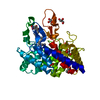
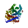
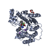
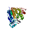
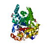
| Title | Glycosidase E178A | ||||||
 Components Components | Glycoside hydrolase 15-related protein | ||||||
 Keywords Keywords |  HYDROLASE / Glycosidase HYDROLASE / Glycosidase | ||||||
| Function / homology | isomaltose glucohydrolase / GH15-like domain / Glycosyl hydrolases family 15 / hydrolase activity, acting on glycosyl bonds / Six-hairpin glycosidase-like superfamily / Six-hairpin glycosidase superfamily / polysaccharide catabolic process / cytoplasm / Isomaltose glucohydrolase Function and homology information Function and homology information | ||||||
| Biological species |  Kribbella flavida DSM 17836 (bacteria) Kribbella flavida DSM 17836 (bacteria) | ||||||
| Method | X-RAY DIFFRACTION / SYNCHROTRON / Resolution: 1.6 Å | ||||||
 Authors Authors | Tanaka, Y. / Chen, M. / Tagami, T. / Yao, M. / Kimura, A. | ||||||
| Funding support |  Japan, 1items Japan, 1items
| ||||||
 Citation Citation | Journal: Febs J. / Year: 2022 Title: Structural insights reveal the second base catalyst of isomaltose glucohydrolase. Authors: Tagami, T. / Chen, M. / Furunaga, Y. / Kikuchi, A. / Sadahiro, J. / Lang, W. / Okuyama, M. / Tanaka, Y. / Iwasaki, T. / Yao, M. / Kimura, A. | ||||||
| History |
|
- Structure visualization
| Structure viewer | Molecule: MolmilJmol/JSmol |
|---|
- Downloads & links
-Download
| PDBx/mmCIF format | 5z3c.cif.gz | 149.6 KB | Display | PDBx/mmCIF format |
|---|---|---|---|---|
| PDB format | pdb5z3c.ent.gz | 123.5 KB | Display | PDB format |
| PDBx/mmJSON format | 5z3c.json.gz | Tree view | PDBx/mmJSON format | |
| Others |  Other downloads Other downloads |
-Validation report
| Arichive directory | https://data.pdbj.org/pub/pdb/validation_reports/z3/5z3cftp://data.pdbj.org/pub/pdb/validation_reports/z3/5z3c | HTTPS FTP |
|---|
-Related structure data
| Related structure data |  5z3aC  5z3bC  5z3dC 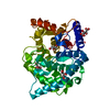 5z3eC  5z3fC  7c24C  7c25C  7c26C  7c27C C: citing same article ( |
|---|---|
| Similar structure data |


-Links
 PDBj
PDBj
- Assembly
| Deposited unit | 
| |||||||||||||||
|---|---|---|---|---|---|---|---|---|---|---|---|---|---|---|---|---|
| 1 |
| |||||||||||||||
| Unit cell |
| |||||||||||||||
| Components on special symmetry positions |
|
-Components
| #1: Protein | Mass: 43970.023 Da / Num. of mol.: 1 / Mutation: E178A Source method: isolated from a genetically manipulated source Source: (gene. exp.) Kribbella flavida DSM 17836 (bacteria) / Strain: DSM 17836 / JCM 10339 / NBRC 14399 / Gene: Kfla_1896 / Production host: Escherichia coli (E. coli) / References: UniProt: D2PPM8 | ||
|---|---|---|---|
| #2: Chemical | ChemComp-GOL / Glycerol  Mass: 92.094 Da / Num. of mol.: 5 / Source method: obtained synthetically / Formula: C3H8O3 Mass: 92.094 Da / Num. of mol.: 5 / Source method: obtained synthetically / Formula: C3H8O3#3: Water | ChemComp-HOH / | Water Mass: 18.015 Da / Num. of mol.: 339 / Source method: isolated from a natural source / Formula: H2O Mass: 18.015 Da / Num. of mol.: 339 / Source method: isolated from a natural source / Formula: H2O |
-Experimental details
-Experiment
| Experiment | Method: X-RAY DIFFRACTION / Number of used crystals: 1 |
|---|
- Sample preparation
| Crystal | Density Matthews: 2.8 Å3/Da / Density % sol: 56.08 % |
|---|---|
| Crystal grow | Temperature: 298 K / Method: vapor diffusion, hanging drop Details: 0.2M Ammonium citrate tribasic (pH 6.8), 0.1M Sodium citrate (pH 5.5), 12% PEGmonomethyl ether 2000, 2mM isomaltose |
-Data collection
| Diffraction | Mean temperature: 100 K |
|---|---|
| Diffraction source | Source: SYNCHROTRON / Site: Photon Factory / Beamline: AR-NE3A / Wavelength: 1 Å |
| Detector | Type: DECTRIS PILATUS 2M / Detector: PIXEL / Date: Jun 4, 2016 |
| Radiation | Protocol: SINGLE WAVELENGTH / Monochromatic (M) / Laue (L): M / Scattering type: x-ray |
| Radiation wavelength | Wavelength: 1 Å / Relative weight: 1 |
| Reflection | Resolution: 1.6→45.26 Å / Num. obs: 66196 / % possible obs: 99.9 % / Redundancy: 21.7 % / Net I/σ(I): 38.12 |
| Reflection shell | Resolution: 1.6→1.69 Å |
- Processing
| Software |
| |||||||||||||||||||||||||||||||||||||||||||||||||||||||||||||||||||||||||||||||||||||||||||||||||||||||||||||||||||||||||||||||||||||||||||||||||||||||||||||||||||||||||||||||
|---|---|---|---|---|---|---|---|---|---|---|---|---|---|---|---|---|---|---|---|---|---|---|---|---|---|---|---|---|---|---|---|---|---|---|---|---|---|---|---|---|---|---|---|---|---|---|---|---|---|---|---|---|---|---|---|---|---|---|---|---|---|---|---|---|---|---|---|---|---|---|---|---|---|---|---|---|---|---|---|---|---|---|---|---|---|---|---|---|---|---|---|---|---|---|---|---|---|---|---|---|---|---|---|---|---|---|---|---|---|---|---|---|---|---|---|---|---|---|---|---|---|---|---|---|---|---|---|---|---|---|---|---|---|---|---|---|---|---|---|---|---|---|---|---|---|---|---|---|---|---|---|---|---|---|---|---|---|---|---|---|---|---|---|---|---|---|---|---|---|---|---|---|---|---|---|---|
| Refinement | Resolution: 1.6→45.257 Å / SU ML: 0.11 / Cross valid method: FREE R-VALUE / σ(F): 1.36 / Phase error: 13.91
| |||||||||||||||||||||||||||||||||||||||||||||||||||||||||||||||||||||||||||||||||||||||||||||||||||||||||||||||||||||||||||||||||||||||||||||||||||||||||||||||||||||||||||||||
| Solvent computation | Shrinkage radii: 0.9 Å / VDW probe radii: 1.11 Å | |||||||||||||||||||||||||||||||||||||||||||||||||||||||||||||||||||||||||||||||||||||||||||||||||||||||||||||||||||||||||||||||||||||||||||||||||||||||||||||||||||||||||||||||
| Refinement step | Cycle: LAST / Resolution: 1.6→45.257 Å
| |||||||||||||||||||||||||||||||||||||||||||||||||||||||||||||||||||||||||||||||||||||||||||||||||||||||||||||||||||||||||||||||||||||||||||||||||||||||||||||||||||||||||||||||
| Refine LS restraints |
| |||||||||||||||||||||||||||||||||||||||||||||||||||||||||||||||||||||||||||||||||||||||||||||||||||||||||||||||||||||||||||||||||||||||||||||||||||||||||||||||||||||||||||||||
| LS refinement shell |
|
