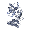
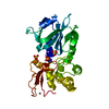
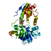
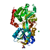
Journal: EMBO Rep / Year: 2019 Title: High-resolution archaellum structure reveals a conserved metal-binding site. Authors: Vladimir A Meshcheryakov / Satoshi Shibata / Makoto Tokoro Schreiber / Alejandro Villar-Briones / Kenneth F Jarrell / Shin-Ichi Aizawa / Matthias Wolf / Abstract: Many archaea swim by means of archaella. While the archaellum is similar in function to its bacterial counterpart, its structure, composition, and evolution are fundamentally different. Archaella are ...Many archaea swim by means of archaella. While the archaellum is similar in function to its bacterial counterpart, its structure, composition, and evolution are fundamentally different. Archaella are related to archaeal and bacterial type IV pili. Despite recent advances, our understanding of molecular processes governing archaellum assembly and stability is still incomplete. Here, we determine the structures of archaella by X-ray crystallography and cryo-EM The crystal structure of FlaB1 is the first and only crystal structure of any archaellin to date at a resolution of 1.5 Å, which is put into biological context by a cryo-EM reconstruction from archaella at 4 Å resolution created with helical single-particle analysis. Our results indicate that the archaellum is predominantly composed of FlaB1. We identify N-linked glycosylation by cryo-EM and mass spectrometry. The crystal structure reveals a highly conserved metal-binding site, which is validated by mass spectrometry and electron energy-loss spectroscopy. We show that the metal-binding site, which appears to be a widespread property of archaellin, is required for filament integrity.
Protocol: SINGLE WAVELENGTH / Monochromatic (M) / Laue (L): M / Scattering type: x-ray
Radiation wavelength
Wavelength: 1 Å / Relative weight: 1
Reflection
Resolution: 1.5→28 Å / Num. obs: 46269 / % possible obs: 96.8 % / Redundancy: 4.3 % / Net I/σ(I): 12.2
Reflection shell
Resolution: 1.5→1.58 Å / Redundancy: 4.3 % / Rmerge(I) obs: 0.325 / Mean I/σ(I) obs: 3.3 / CC1/2: 0.883 / % possible all: 92.9
-
Processing
Software
Name
Version
Classification
REFMAC
5.8.0107
refinement
MOSFLM
datareduction
SCALA
datascaling
PHENIX
phasing
Refinement
Method to determine structure: MIR / Resolution: 1.5→28 Å / Cor.coef. Fo:Fc: 0.966 / Cor.coef. Fo:Fc free: 0.951 / SU B: 1.547 / SU ML: 0.056 / Cross valid method: THROUGHOUT / ESU R: 0.076 / ESU R Free: 0.077 / Details: HYDROGENS HAVE BEEN ADDED IN THE RIDING POSITIONS
Rfactor
Num. reflection
% reflection
Selection details
Rfree
0.2
2283
4.9 %
RANDOM
Rwork
0.17273
-
-
-
obs
0.17408
43943
96.36 %
-
Solvent computation
Ion probe radii: 0.8 Å / Shrinkage radii: 0.8 Å / VDW probe radii: 1.2 Å
 Movie
Movie Controller

 Yorodumi
 Open data
 Basic information
 Components
 Keywords
 STRUCTURAL PROTEIN /
  Function and homology information

 Authors
 Citation

 Structure visualization
 Downloads & links
 Other downloads
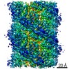


 PDBj
PDBj Assembly
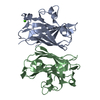


 Mass: 40.078 Da / Num. of mol.: 2 / Source method: obtained synthetically / Formula: Ca
 Mass: 18.015 Da / Num. of mol.: 248 / Source method: isolated from a natural source / Formula: H2O
 Sample preparation
 Processing