 Movie
Movie Controller

[English] 日本語
 Yorodumi
- PDB-5y04: Crystal Structure of the complex between the vinculin D1 domain a... -
+ Open data

- Basic information
| Entry | Database: PDB / ID: 5y04 | ||||||
|---|---|---|---|---|---|---|---|
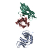
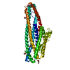
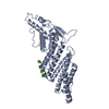
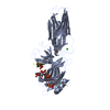
| Title | Crystal Structure of the complex between the vinculin D1 domain and alphaE-catenin | ||||||
 Components Components |
| ||||||
 Keywords Keywords |  CELL ADHESION / ADHERENS JUNCTION / CYTOSKELETON CELL ADHESION / ADHERENS JUNCTION / CYTOSKELETON | ||||||
| Function / homology |  Function and homology information Function and homology informationsmall GTPase binding => GO:0031267 / negative regulation of integrin-mediated signaling pathway / VEGFR2 mediated vascular permeability / regulation of protein localization to adherens junction / outer dense plaque of desmosome / inner dense plaque of desmosome / Smooth Muscle Contraction / MAP2K and MAPK activation / terminal web / RHO GTPases activate IQGAPs ...small GTPase binding => GO:0031267 / negative regulation of integrin-mediated signaling pathway / VEGFR2 mediated vascular permeability / regulation of protein localization to adherens junction / outer dense plaque of desmosome / inner dense plaque of desmosome / Smooth Muscle Contraction / MAP2K and MAPK activation / terminal web / RHO GTPases activate IQGAPs / Adherens junctions interactions / gap junction assembly / epithelial cell-cell adhesion / zonula adherens / Platelet degranulation / dystroglycan binding / gamma-catenin binding / vinculin binding / alpha-catenin binding / cellular response to indole-3-methanol / flotillin complex / fascia adherens / negative regulation of cell motility / cell-cell contact zone / Myogenesis / costamere / apical junction assembly / regulation of establishment of endothelial barrier / adherens junction assembly / positive regulation of extrinsic apoptotic signaling pathway in absence of ligand / positive regulation of smoothened signaling pathway / catenin complex / axon extension / protein localization to cell surface / podosome / negative regulation of protein localization to nucleus / lamellipodium assembly / axon regeneration / negative regulation of neuroblast proliferation / regulation of focal adhesion assembly / smoothened signaling pathway / establishment or maintenance of cell polarity / odontogenesis of dentin-containing tooth / brush border / neuroblast proliferation / negative regulation of extrinsic apoptotic signaling pathway in absence of ligand / intercalated disc / regulation of cell migration / ovarian follicle development / extrinsic apoptotic signaling pathway in absence of ligand / Neutrophil degranulation / acrosomal vesicle / cell motility / integrin-mediated signaling pathway / morphogenesis of an epithelium / adherens junction / sarcolemma / protein localization / cell-cell adhesion / Z disc / beta-catenin binding / response to estrogen / male gonad development / actin filament binding / cell-cell junction / cell migration / actin cytoskeleton / lamellipodium / cell junction / regulation of cell population proliferation / cell adhesion / cadherin binding / membrane raft / focal adhesion / intracellular membrane-bounded organelle / apoptotic process / ubiquitin protein ligase binding / protein-containing complex binding / structural molecule activity / negative regulation of apoptotic process / protein-containing complex / identical protein binding / plasma membrane / cytoplasmSimilarity search - Function | ||||||
| Biological species |  Mus musculus (house mouse) Mus musculus (house mouse) | ||||||
| Method | X-RAY DIFFRACTION / SYNCHROTRON / MAD / Resolution: 2.85 Å | ||||||
 Authors Authors | Hirano, Y. / Hakoshima, T. | ||||||
 Citation Citation | Journal: Genes Cells / Year: 2018 Title: The force-sensing device region of alpha-catenin is an intrinsically disordered segment in the absence of intramolecular stabilization of the autoinhibitory form Authors: Hirano, Y. / Amano, Y. / Yonemura, S. / Hakoshima, T. | ||||||
| History |
|
- Structure visualization
| Structure viewer | Molecule: MolmilJmol/JSmol |
|---|
- Downloads & links
-Download
| PDBx/mmCIF format | 5y04.cif.gz | 126.7 KB | Display | PDBx/mmCIF format |
|---|---|---|---|---|
| PDB format | pdb5y04.ent.gz | 98.8 KB | Display | PDB format |
| PDBx/mmJSON format | 5y04.json.gz | Tree view | PDBx/mmJSON format | |
| Others |  Other downloads Other downloads |
-Validation report
| Arichive directory | https://data.pdbj.org/pub/pdb/validation_reports/y0/5y04ftp://data.pdbj.org/pub/pdb/validation_reports/y0/5y04 | HTTPS FTP |
|---|
-Related structure data
| Related structure data |  5xflC  3w3r C: citing same article ( |
|---|---|
| Similar structure data |


-Links
 PDBj
PDBj


- Assembly
| Deposited unit | 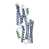
| ||||||||
|---|---|---|---|---|---|---|---|---|---|
| 1 |
| ||||||||
| Unit cell |
|
-Components
| #1: Protein | / Metavinculin Mass: 28100.598 Da / Num. of mol.: 1 / Fragment: UNP RESIDUES 1-250 Source method: isolated from a genetically manipulated source Source: (gene. exp.) Mus musculus (house mouse) / Gene: Vcl / Plasmid: PET49B / Production host:  Escherichia coli (E. coli) / Strain (production host): BL21 STAR (DE3) / References: UniProt: Q64727 Escherichia coli (E. coli) / Strain (production host): BL21 STAR (DE3) / References: UniProt: Q64727 |
|---|---|
| #2: Protein | / 102 kDa cadherin-associated protein / Alpha E-catenin / CAP102 Mass: 11410.797 Da / Num. of mol.: 1 / Fragment: UNP RESIDUES 276-375 Source method: isolated from a genetically manipulated source Source: (gene. exp.) Mus musculus (house mouse) / Gene: Ctnna1, Catna1 / Plasmid: PET49B / Production host: Escherichia coli (E. coli) / Strain (production host): BL21 STAR (DE3) / References: UniProt: P26231 |
| #3: Water | ChemComp-HOH / Water Mass: 18.015 Da / Num. of mol.: 6 / Source method: isolated from a natural source / Formula: H2O Mass: 18.015 Da / Num. of mol.: 6 / Source method: isolated from a natural source / Formula: H2O |
-Experimental details
-Experiment
| Experiment | Method: X-RAY DIFFRACTION / Number of used crystals: 1 |
|---|
- Sample preparation
| Crystal | Density Matthews: 2.85 Å3/Da / Density % sol: 56.9 % |
|---|---|
| Crystal grow | Temperature: 293 K / Method: vapor diffusion / pH: 8 Details: 100MM IMIDAZOLE, 0.5-0.7M POTASSIUM TARTRATE, 0.35M SODIUM FORMATE PH range: 8 |
-Data collection
| Diffraction | Mean temperature: 100 K | ||||||||||||
|---|---|---|---|---|---|---|---|---|---|---|---|---|---|
| Diffraction source | Source: SYNCHROTRON / Site: SPring-8  / Beamline: BL41XU / Wavelength: 0.9785, 0.9788, 0.9946 / Beamline: BL41XU / Wavelength: 0.9785, 0.9788, 0.9946 | ||||||||||||
| Detector | Type: RAYONIX MX225HE / Detector: CCD / Date: Oct 19, 2009 / Details: MIRRORS | ||||||||||||
| Radiation | Monochromator: ROTATED-INCLINED DOUBLE-CRYSTAL MONOCHROMATOR , SI (111) Protocol: MAD / Monochromatic (M) / Laue (L): M / Scattering type: x-ray | ||||||||||||
| Radiation wavelength |
| ||||||||||||
| Reflection | Resolution: 2.85→50 Å / Num. obs: 10287 / % possible obs: 96 % / Observed criterion σ(I): 0 / Redundancy: 6.7 % / Rsym value: 0.052 / Net I/σ(I): 41.4 | ||||||||||||
| Reflection shell | Resolution: 2.85→2.95 Å / Redundancy: 4.2 % / Mean I/σ(I) obs: 2.8 / Rsym value: 0.425 / % possible all: 73.4 |
- Processing
| Software |
| ||||||||||||||||||||||||||||||||||||||||||||||||||||||||||||||||||||||||||||||||||||||||||||||||||||||||||||||||||||||||||||||||||||||||||||||||||||||||||||||||||||||||||||||||||||||
|---|---|---|---|---|---|---|---|---|---|---|---|---|---|---|---|---|---|---|---|---|---|---|---|---|---|---|---|---|---|---|---|---|---|---|---|---|---|---|---|---|---|---|---|---|---|---|---|---|---|---|---|---|---|---|---|---|---|---|---|---|---|---|---|---|---|---|---|---|---|---|---|---|---|---|---|---|---|---|---|---|---|---|---|---|---|---|---|---|---|---|---|---|---|---|---|---|---|---|---|---|---|---|---|---|---|---|---|---|---|---|---|---|---|---|---|---|---|---|---|---|---|---|---|---|---|---|---|---|---|---|---|---|---|---|---|---|---|---|---|---|---|---|---|---|---|---|---|---|---|---|---|---|---|---|---|---|---|---|---|---|---|---|---|---|---|---|---|---|---|---|---|---|---|---|---|---|---|---|---|---|---|---|---|
| Refinement | Method to determine structure: MAD / Resolution: 2.85→50 Å / Cor.coef. Fo:Fc: 0.936 / Cor.coef. Fo:Fc free: 0.915 / SU B: 34.624 / SU ML: 0.293 / Cross valid method: THROUGHOUT / σ(F): 0 / ESU R: 1.053 / ESU R Free: 0.392 / Details: HYDROGENS HAVE BEEN USED IF PRESENT IN THE INPUT
| ||||||||||||||||||||||||||||||||||||||||||||||||||||||||||||||||||||||||||||||||||||||||||||||||||||||||||||||||||||||||||||||||||||||||||||||||||||||||||||||||||||||||||||||||||||||
| Solvent computation | Ion probe radii: 0.8 Å / Shrinkage radii: 0.8 Å / VDW probe radii: 1.2 Å | ||||||||||||||||||||||||||||||||||||||||||||||||||||||||||||||||||||||||||||||||||||||||||||||||||||||||||||||||||||||||||||||||||||||||||||||||||||||||||||||||||||||||||||||||||||||
| Displacement parameters | Biso mean: 94 Å2
| ||||||||||||||||||||||||||||||||||||||||||||||||||||||||||||||||||||||||||||||||||||||||||||||||||||||||||||||||||||||||||||||||||||||||||||||||||||||||||||||||||||||||||||||||||||||
| Refinement step | Cycle: LAST / Resolution: 2.85→50 Å
| ||||||||||||||||||||||||||||||||||||||||||||||||||||||||||||||||||||||||||||||||||||||||||||||||||||||||||||||||||||||||||||||||||||||||||||||||||||||||||||||||||||||||||||||||||||||
| Refine LS restraints |
|