 Movie
Movie Controller

[English] 日本語
 Yorodumi
- PDB-5xf1: Structure of the Full-length glucagon class B G protein-coupled r... -
+ Open data

- Basic information
| Entry | Database: PDB / ID: 5xf1 | |||||||||
|---|---|---|---|---|---|---|---|---|---|---|
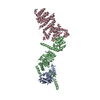
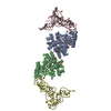
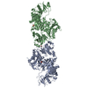
| Title | Structure of the Full-length glucagon class B G protein-coupled receptor | |||||||||
 Components Components |
| |||||||||
 Keywords Keywords |  SIGNALING PROTEIN / Human GCGR receptor / Class B / 7TM domain / membrane / LCP / XFEL SIGNALING PROTEIN / Human GCGR receptor / Class B / 7TM domain / membrane / LCP / XFEL | |||||||||
| Function / homology |  Function and homology information Function and homology informationregulation of glycogen metabolic process / glucagon receptor activity / response to starvation / cellular response to glucagon stimulus / exocytosis / peptide hormone binding / viral release from host cell by cytolysis / cellular response to starvation / hormone-mediated signaling pathway / guanyl-nucleotide exchange factor activity ...regulation of glycogen metabolic process / glucagon receptor activity / response to starvation / cellular response to glucagon stimulus / exocytosis / peptide hormone binding / viral release from host cell by cytolysis / cellular response to starvation / hormone-mediated signaling pathway / guanyl-nucleotide exchange factor activity / peptidoglycan catabolic process / response to nutrient / generation of precursor metabolites and energy / adenylate cyclase-modulating G protein-coupled receptor signaling pathway / Glucagon signaling in metabolic regulation / adenylate cyclase-activating G protein-coupled receptor signaling pathway / Glucagon-type ligand receptors / regulation of blood pressure / cell wall macromolecule catabolic process / glucose homeostasis / lysozyme / lysozyme activity / G alpha (s) signalling events / G alpha (q) signalling events / host cell cytoplasm / cell surface receptor signaling pathway / endosome / defense response to bacterium / positive regulation of gene expression / membrane / plasma membraneSimilarity search - Function | |||||||||
| Biological species |  Homo sapiens (human) Homo sapiens (human) Enterobacteria phage T4 (virus) Enterobacteria phage T4 (virus) | |||||||||
| Method | X-RAY DIFFRACTION / SYNCHROTRON / MOLECULAR REPLACEMENT / Resolution: 3.19 Å | |||||||||
 Authors Authors | Zhang, H. / Qiao, A. / Yang, D. / Yang, L. / Dai, A. / de Graaf, C. / Reedtz-Runge, S. / Dharmarajan, V. / Zhang, H. / Han, G.W. ...Zhang, H. / Qiao, A. / Yang, D. / Yang, L. / Dai, A. / de Graaf, C. / Reedtz-Runge, S. / Dharmarajan, V. / Zhang, H. / Han, G.W. / Grant, T. / Sierra, R. / Weierstall, U. / Nelson, G. / Liu, W. / Wu, Y. / Ma, L. / Cai, X. / Lin, G. / Wu, X. / Geng, Z. / Dong, Y. / Song, G. / Griffin, P. / Lau, J. / Cherezov, V. / Yang, H. / Hanson, M. / Stevens, R. / Jiang, H. / Wang, M. / Zhao, Q. / Wu, B. | |||||||||
 Citation Citation | Journal: Nature / Year: 2017 Title: Structure of the full-length glucagon class B G-protein-coupled receptor. Authors: Zhang, H. / Qiao, A. / Yang, D. / Yang, L. / Dai, A. / de Graaf, C. / Reedtz-Runge, S. / Dharmarajan, V. / Zhang, H. / Han, G.W. / Grant, T.D. / Sierra, R.G. / Weierstall, U. / Nelson, G. / ...Authors: Zhang, H. / Qiao, A. / Yang, D. / Yang, L. / Dai, A. / de Graaf, C. / Reedtz-Runge, S. / Dharmarajan, V. / Zhang, H. / Han, G.W. / Grant, T.D. / Sierra, R.G. / Weierstall, U. / Nelson, G. / Liu, W. / Wu, Y. / Ma, L. / Cai, X. / Lin, G. / Wu, X. / Geng, Z. / Dong, Y. / Song, G. / Griffin, P.R. / Lau, J. / Cherezov, V. / Yang, H. / Hanson, M.A. / Stevens, R.C. / Zhao, Q. / Jiang, H. / Wang, M.W. / Wu, B. | |||||||||
| History |
|
- Structure visualization
| Structure viewer | Molecule: MolmilJmol/JSmol |
|---|
- Downloads & links
-Download
| PDBx/mmCIF format | 5xf1.cif.gz | 726.6 KB | Display | PDBx/mmCIF format |
|---|---|---|---|---|
| PDB format | pdb5xf1.ent.gz | 603.5 KB | Display | PDB format |
| PDBx/mmJSON format | 5xf1.json.gz | Tree view | PDBx/mmJSON format | |
| Others |  Other downloads Other downloads |
-Validation report
| Arichive directory | https://data.pdbj.org/pub/pdb/validation_reports/xf/5xf1ftp://data.pdbj.org/pub/pdb/validation_reports/xf/5xf1 | HTTPS FTP |
|---|
-Related structure data
| Related structure data |  5xezC  4l6rS C: citing same article ( S: Starting model for refinement |
|---|---|
| Similar structure data |


-Links
 PDBj
PDBj


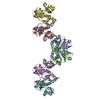
- Assembly
| Deposited unit | 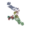
| |||||||||||||||||||||||||||||||||||||||||||||||||||||||||||||||||||
|---|---|---|---|---|---|---|---|---|---|---|---|---|---|---|---|---|---|---|---|---|---|---|---|---|---|---|---|---|---|---|---|---|---|---|---|---|---|---|---|---|---|---|---|---|---|---|---|---|---|---|---|---|---|---|---|---|---|---|---|---|---|---|---|---|---|---|---|---|
| 1 | 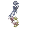
| |||||||||||||||||||||||||||||||||||||||||||||||||||||||||||||||||||
| 2 | 
| |||||||||||||||||||||||||||||||||||||||||||||||||||||||||||||||||||
| Unit cell |
| |||||||||||||||||||||||||||||||||||||||||||||||||||||||||||||||||||
| Noncrystallographic symmetry (NCS) | NCS domain:
NCS domain segments:
NCS ensembles :
|
-Components
-Protein , 1 types, 2 molecules AB
| #1: Protein | Mass: 65784.070 Da / Num. of mol.: 2 Fragment: UNP RESIDUES 27-256,UNP RESIDUES 2-161,UNP RESIDUES 260-432 Mutation: C54T, C97A Source method: isolated from a genetically manipulated source Details: The fusion protein of Glucagon receptor (UNP RESIDUES 27-256), Endolysin (UNP RESIDUES 2-161) and Glucagon receptor (UNP RESIDUES 260-432) Source: (gene. exp.) Homo sapiens (human), (gene. exp.) Enterobacteria phage T4 (virus)Gene: GCGR, e, T4Tp126 / Production host:   Spodoptera frugiperda (fall armyworm) / References: UniProt: P47871, UniProt: D9IEF7, lysozyme Spodoptera frugiperda (fall armyworm) / References: UniProt: P47871, UniProt: D9IEF7, lysozyme |
|---|
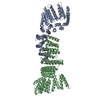
-Antibody , 2 types, 4 molecules CHDL
| #2: Antibody | Mass: 24977.961 Da / Num. of mol.: 2 Source method: isolated from a genetically manipulated source Source: (gene. exp.) Homo sapiens (human) / Cell line (production host): HEK293 / Production host: Homo sapiens (human)#3: Antibody | Mass: 23263.811 Da / Num. of mol.: 2 Source method: isolated from a genetically manipulated source Source: (gene. exp.) Homo sapiens (human) / Cell line (production host): HEK293 / Production host: Homo sapiens (human) |
|---|
-Sugars , 3 types, 7 molecules 
| #4: Polysaccharide | 2-acetamido-2-deoxy-beta-D-glucopyranose-(1-4)-2-acetamido-2-deoxy-beta-D-glucopyranose / Mass: 424.401 Da / Num. of mol.: 5Source method: isolated from a genetically manipulated source #5: Polysaccharide | 2-acetamido-2-deoxy-beta-D-glucopyranose-(1-4)-2-acetamido-2-deoxy-beta-D-glucopyranose-(1-4)-2- ...2-acetamido-2-deoxy-beta-D-glucopyranose-(1-4)-2-acetamido-2-deoxy-beta-D-glucopyranose-(1-4)-2-acetamido-2-deoxy-beta-D-glucopyranose / triacetyl-beta-chitotriose |   , Oligosaccharide / Class: Inhibitor / Mass: 627.594 Da / Num. of mol.: 1 , Oligosaccharide / Class: Inhibitor / Mass: 627.594 Da / Num. of mol.: 1Source method: isolated from a genetically manipulated source Details: oligosaccharide / References: triacetyl-beta-chitotriose #7: Sugar | ChemComp-NAG / | N-Acetylglucosamine Type: D-saccharide, beta linking / Mass: 221.208 Da / Num. of mol.: 1 Type: D-saccharide, beta linking / Mass: 221.208 Da / Num. of mol.: 1Source method: isolated from a genetically manipulated source Formula: C8H15NO6 |
|---|
-Non-polymers , 2 types, 3 molecules 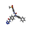

| #6: Chemical |  Mass: 573.666 Da / Num. of mol.: 2 / Source method: obtained synthetically / Formula: C29H31N7O4S Mass: 573.666 Da / Num. of mol.: 2 / Source method: obtained synthetically / Formula: C29H31N7O4S#8: Water | ChemComp-HOH / | WaterMass: 18.015 Da / Num. of mol.: 1 / Source method: isolated from a natural source / Formula: H2O |
|---|
-Experimental details
-Experiment
| Experiment | Method: X-RAY DIFFRACTION / Number of used crystals: 1 |
|---|
- Sample preparation
| Crystal | Density Matthews: 3.68 Å3/Da / Density % sol: 66.56 % |
|---|---|
| Crystal grow | Temperature: 293 K / Method: lipidic cubic phase Details: 100mM HEPES, pH7.0, 200mM potassium phospphate monobasic, 20% PEG500DME, 10 mM gly-gly-glysine |
-Data collection
| Diffraction | Mean temperature: 100 K | |||||||||||||||
|---|---|---|---|---|---|---|---|---|---|---|---|---|---|---|---|---|
| Diffraction source | Source: SYNCHROTRON / Site: SPring-8  / Beamline: BL41XU / Wavelength: 1 Å / Beamline: BL41XU / Wavelength: 1 Å | |||||||||||||||
| Detector | Type: DECTRIS PILATUS3 S 6M / Detector: PIXEL / Date: Jul 20, 2016 | |||||||||||||||
| Radiation | Protocol: SINGLE WAVELENGTH / Monochromatic (M) / Laue (L): M / Scattering type: x-ray | |||||||||||||||
| Radiation wavelength | Wavelength: 1 Å / Relative weight: 1 | |||||||||||||||
| Reflection twin |
| |||||||||||||||
| Reflection | Resolution: 3.19→50 Å / Num. obs: 49677 / % possible obs: 92.8 % / Redundancy: 2.8 % / Net I/σ(I): 3.1 |
- Processing
| Software |
| ||||||||||||||||||||||||||||||||||||||||||||||||||||||||||||||||||||||||||||||||||||||||||||||||||||||||||||||||||||||||||||||||||||||||||||||||||||||||||||||||||||||||||||||||||||||
|---|---|---|---|---|---|---|---|---|---|---|---|---|---|---|---|---|---|---|---|---|---|---|---|---|---|---|---|---|---|---|---|---|---|---|---|---|---|---|---|---|---|---|---|---|---|---|---|---|---|---|---|---|---|---|---|---|---|---|---|---|---|---|---|---|---|---|---|---|---|---|---|---|---|---|---|---|---|---|---|---|---|---|---|---|---|---|---|---|---|---|---|---|---|---|---|---|---|---|---|---|---|---|---|---|---|---|---|---|---|---|---|---|---|---|---|---|---|---|---|---|---|---|---|---|---|---|---|---|---|---|---|---|---|---|---|---|---|---|---|---|---|---|---|---|---|---|---|---|---|---|---|---|---|---|---|---|---|---|---|---|---|---|---|---|---|---|---|---|---|---|---|---|---|---|---|---|---|---|---|---|---|---|---|
| Refinement | Method to determine structure: MOLECULAR REPLACEMENT Starting model: 4L6R Resolution: 3.19→46.92 Å / Cor.coef. Fo:Fc: 0.922 / Cor.coef. Fo:Fc free: 0.879 / SU B: 19.837 / SU ML: 0.165 / Cross valid method: THROUGHOUT / ESU R Free: 0.091 / Stereochemistry target values: MAXIMUM LIKELIHOOD / Details: HYDROGENS HAVE BEEN ADDED IN THE RIDING POSITIONS
| ||||||||||||||||||||||||||||||||||||||||||||||||||||||||||||||||||||||||||||||||||||||||||||||||||||||||||||||||||||||||||||||||||||||||||||||||||||||||||||||||||||||||||||||||||||||
| Solvent computation | Ion probe radii: 0.8 Å / Shrinkage radii: 0.8 Å / VDW probe radii: 1.2 Å / Solvent model: MASK | ||||||||||||||||||||||||||||||||||||||||||||||||||||||||||||||||||||||||||||||||||||||||||||||||||||||||||||||||||||||||||||||||||||||||||||||||||||||||||||||||||||||||||||||||||||||
| Displacement parameters | Biso mean: 116.895 Å2
| ||||||||||||||||||||||||||||||||||||||||||||||||||||||||||||||||||||||||||||||||||||||||||||||||||||||||||||||||||||||||||||||||||||||||||||||||||||||||||||||||||||||||||||||||||||||
| Refinement step | Cycle: 1 / Resolution: 3.19→46.92 Å
| ||||||||||||||||||||||||||||||||||||||||||||||||||||||||||||||||||||||||||||||||||||||||||||||||||||||||||||||||||||||||||||||||||||||||||||||||||||||||||||||||||||||||||||||||||||||
| Refine LS restraints |
|