 Movie
Movie Controller

[English] 日本語
 Yorodumi
- PDB-5wwz: Crystal structure of the KH2 domain of human RNA-binding E3 ubiqu... -
+ Open data

- Basic information
| Entry | Database: PDB / ID: 5wwz | ||||||
|---|---|---|---|---|---|---|---|
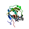
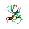
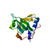
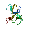
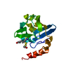
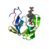
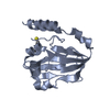
| Title | Crystal structure of the KH2 domain of human RNA-binding E3 ubiquitin-protein ligase MEX-3C | ||||||
 Components Components | RNA-binding E3 ubiquitin-protein ligase MEX3C | ||||||
 Keywords Keywords |  RNA BINDING PROTEIN / KH2 / MEX-3C RNA BINDING PROTEIN / KH2 / MEX-3C | ||||||
| Function / homology |  Function and homology information Function and homology informationchondrocyte hypertrophy / regulation of fat cell differentiation / energy homeostasis / RING-type E3 ubiquitin transferase / ubiquitin protein ligase activity / Antigen processing: Ubiquitination & Proteasome degradation / RNA binding / metal ion binding / nucleus / cytoplasmSimilarity search - Function | ||||||
| Biological species |  Homo sapiens (human) Homo sapiens (human) | ||||||
| Method | X-RAY DIFFRACTION / SYNCHROTRON / MOLECULAR REPLACEMENT / Resolution: 2.5 Å | ||||||
 Authors Authors | Yang, L. / Wang, C. / Li, F. / Gong, Q. | ||||||
 Citation Citation | Journal: J. Biol. Chem. / Year: 2017 Title: The human RNA-binding protein and E3 ligase MEX-3C binds the MEX-3-recognition element (MRE) motif with high affinity Authors: Yang, L. / Wang, C. / Li, F. / Zhang, J. / Nayab, A. / Wu, J. / Shi, Y. / Gong, Q. | ||||||
| History |
|
- Structure visualization
| Structure viewer | Molecule: MolmilJmol/JSmol |
|---|
- Downloads & links
-Download
| PDBx/mmCIF format | 5wwz.cif.gz | 106.6 KB | Display | PDBx/mmCIF format |
|---|---|---|---|---|
| PDB format | pdb5wwz.ent.gz | 81.7 KB | Display | PDB format |
| PDBx/mmJSON format | 5wwz.json.gz | Tree view | PDBx/mmJSON format | |
| Others |  Other downloads Other downloads |
-Validation report
| Arichive directory | https://data.pdbj.org/pub/pdb/validation_reports/ww/5wwzftp://data.pdbj.org/pub/pdb/validation_reports/ww/5wwz | HTTPS FTP |
|---|
-Related structure data
| Related structure data |  5wwwSC  5wwxC S: Starting model for refinement C: citing same article ( |
|---|---|
| Similar structure data |


-Links
 PDBj
PDBj


- Assembly
| Deposited unit | 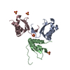
| ||||||||||||||||||||||||||||||||||||||||||||||||||||||||||||||||||||||||||||
|---|---|---|---|---|---|---|---|---|---|---|---|---|---|---|---|---|---|---|---|---|---|---|---|---|---|---|---|---|---|---|---|---|---|---|---|---|---|---|---|---|---|---|---|---|---|---|---|---|---|---|---|---|---|---|---|---|---|---|---|---|---|---|---|---|---|---|---|---|---|---|---|---|---|---|---|---|---|
| 1 | 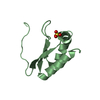
| ||||||||||||||||||||||||||||||||||||||||||||||||||||||||||||||||||||||||||||
| 2 | 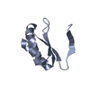
| ||||||||||||||||||||||||||||||||||||||||||||||||||||||||||||||||||||||||||||
| 3 | 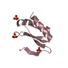
| ||||||||||||||||||||||||||||||||||||||||||||||||||||||||||||||||||||||||||||
| Unit cell |
| ||||||||||||||||||||||||||||||||||||||||||||||||||||||||||||||||||||||||||||
| Noncrystallographic symmetry (NCS) | NCS domain:
NCS domain segments:
|