 Movie
Movie Controller


+ Open data

- Basic information
| Entry | Database: PDB / ID: 5wqv | ||||||
|---|---|---|---|---|---|---|---|
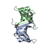
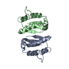
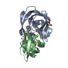
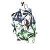
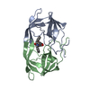
| Title | Crystal structure of PriB mutant - S55A | ||||||
 Components Components | Primosomal replication protein N | ||||||
 Keywords Keywords |  DNA BINDING PROTEIN / Bacterial DNA replication / OB-fold / S55A mutant DNA BINDING PROTEIN / Bacterial DNA replication / OB-fold / S55A mutant | ||||||
| Function / homology |  Function and homology information Function and homology informationpre-primosome complex / DnaB-DnaC-DnaT-PriA-PriB complex / plasmid maintenance / primosome complex / DNA replication, synthesis of primer / replication fork processing / DNA unwinding involved in DNA replication / DNA replication initiation / response to radiation / single-stranded DNA binding / identical protein bindingSimilarity search - Function | ||||||
| Biological species |  Escherichia coli (E. coli) Escherichia coli (E. coli) | ||||||
| Method | X-RAY DIFFRACTION / SYNCHROTRON / MOLECULAR REPLACEMENT / Resolution: 1.97 Å | ||||||
 Authors Authors | Fujiyama, S. / Shiroishi, M. / Katayama, T. / Abe, Y. / Ueda, T. | ||||||
| Funding support |  Japan, 1items Japan, 1items
| ||||||
 Citation Citation | Journal: Biochim Biophys Acta Proteins Proteom / Year: 2019 Title: Insight into the interaction between PriB and DnaT on bacterial DNA replication restart: Significance of the residues on PriB dimer interface and highly acidic region on DnaT. Authors: Fujiyama, S. / Abe, Y. / Shiroishi, M. / Ikeda, Y. / Ueda, T. | ||||||
| History |
|
- Structure visualization
| Structure viewer | Molecule: MolmilJmol/JSmol |
|---|
- Downloads & links
-Download
| PDBx/mmCIF format | 5wqv.cif.gz | 79.7 KB | Display | PDBx/mmCIF format |
|---|---|---|---|---|
| PDB format | pdb5wqv.ent.gz | 62.7 KB | Display | PDB format |
| PDBx/mmJSON format | 5wqv.json.gz | Tree view | PDBx/mmJSON format | |
| Others |  Other downloads Other downloads |
-Validation report
| Arichive directory | https://data.pdbj.org/pub/pdb/validation_reports/wq/5wqvftp://data.pdbj.org/pub/pdb/validation_reports/wq/5wqv | HTTPS FTP |
|---|
-Related structure data
| Similar structure data |
|---|


-Links
 PDBj
PDBj- Assembly
| Deposited unit | 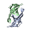
| ||||||||
|---|---|---|---|---|---|---|---|---|---|
| 1 |
| ||||||||
| Unit cell |
|
-Components
| #1: Protein | Mass: 11443.194 Da / Num. of mol.: 2 / Mutation: S55A Source method: isolated from a genetically manipulated source Source: (gene. exp.) Escherichia coli (strain K12) (bacteria)Strain: K12 / Gene: priB, BWG_3913 / Plasmid: pET22b / Production host: Escherichia coli BL21(DE3) (bacteria) / Strain (production host): BL21(DE3) / References: UniProt: C4ZR78, UniProt: P07013*PLUS#2: Water | ChemComp-HOH / | Water Mass: 18.015 Da / Num. of mol.: 97 / Source method: isolated from a natural source / Formula: H2O Mass: 18.015 Da / Num. of mol.: 97 / Source method: isolated from a natural source / Formula: H2O |
|---|
-Experimental details
-Experiment
| Experiment | Method: X-RAY DIFFRACTION / Number of used crystals: 1 |
|---|
- Sample preparation
| Crystal | Density Matthews: 2.1 Å3/Da / Density % sol: 41.6 % |
|---|---|
| Crystal grow | Temperature: 293 K / Method: vapor diffusion, sitting drop / pH: 7 / Details: HEPES, PEG 400, ammonium sulfate |
-Data collection
| Diffraction | Mean temperature: 100 K |
|---|---|
| Diffraction source | Source: SYNCHROTRON / Site: SPring-8 / Beamline: BL38B1 / Wavelength: 1 Å |
| Detector | Type: MARMOSAIC 225 mm CCD / Detector: CCD / Date: Apr 21, 2016 |
| Radiation | Protocol: SINGLE WAVELENGTH / Monochromatic (M) / Laue (L): M / Scattering type: x-ray |
| Radiation wavelength | Wavelength: 1 Å / Relative weight: 1 |
| Reflection | Resolution: 1.97→50 Å / Num. obs: 13685 / % possible obs: 96.8 % / Redundancy: 6.9 % / Biso Wilson estimate: 26 Å2 / Rmerge(I) obs: 0.093 / Net I/σ(I): 35.3 |
| Reflection shell | Resolution: 1.97→2 Å / Redundancy: 7.1 % / Rmerge(I) obs: 0.465 / Mean I/σ(I) obs: 5.2 / % possible all: 99.9 |
- Processing
| Software |
| ||||||||||||||||||||
|---|---|---|---|---|---|---|---|---|---|---|---|---|---|---|---|---|---|---|---|---|---|
| Refinement | Method to determine structure: MOLECULAR REPLACEMENT / Resolution: 1.97→39.6 Å / Cross valid method: FREE R-VALUE
| ||||||||||||||||||||
| Displacement parameters | Biso mean: 28 Å2 | ||||||||||||||||||||
| Refinement step | Cycle: LAST / Resolution: 1.97→39.6 Å
| ||||||||||||||||||||
| LS refinement shell | Resolution: 1.97→2.12 Å
|
