National Institutes of Health/National Institute of General Medical Sciences (NIH/NIGMS)
GM43949
United States
Howard Hughes Medical Institute (HHMI)
United States
Citation
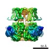
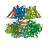
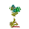
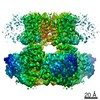
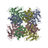
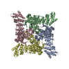
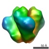
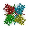
Journal: Nature / Year: 2017 Title: Cryo-EM structure of the open high-conductance Ca-activated K channel. Authors: Xiao Tao / Richard K Hite / Roderick MacKinnon / Abstract: The Ca-activated K channel, Slo1, has an unusually large conductance and contains a voltage sensor and multiple chemical sensors. Dual activation by membrane voltage and Ca renders Slo1 central to ...The Ca-activated K channel, Slo1, has an unusually large conductance and contains a voltage sensor and multiple chemical sensors. Dual activation by membrane voltage and Ca renders Slo1 central to processes that couple electrical signalling to Ca-mediated events such as muscle contraction and neuronal excitability. Here we present the cryo-electron microscopy structure of a full-length Slo1 channel from Aplysia californica in the presence of Ca and Mg at a resolution of 3.5 Å. The channel adopts an open conformation. Its voltage-sensor domain adopts a non-domain-swapped attachment to the pore and contacts the cytoplasmic Ca-binding domain from a neighbouring subunit. Unique structural features of the Slo1 voltage sensor suggest that it undergoes different conformational changes than other known voltage sensors. The structure reveals the molecular details of three distinct divalent cation-binding sites identified through electrophysiological studies of mutant Slo1 channels.
Average exposure time: 0.3 sec. / Electron dose: 1.8 e/Å2 / Detector mode: SUPER-RESOLUTION / Film or detector model: GATAN K2 SUMMIT (4k x 4k) / Num. of grids imaged: 1 / Num. of real images: 2000
Image scans
Sampling size: 5 µm / Width: 7420 / Height: 7676 / Movie frames/image: 50 / Used frames/image: 1-50
Resolution: 3.5 Å / Resolution method: FSC 0.143 CUT-OFF / Num. of particles: 115000 / Symmetry type: POINT
Atomic model building
B value: 100 / Protocol: AB INITIO MODEL / Space: RECIPROCAL
Refinement
Resolution: 3.5→3.5 Å / Cor.coef. Fo:Fc: 0.85 / SU B: 39.858 / SU ML: 0.48 / ESU R: 1.568 Stereochemistry target values: MAXIMUM LIKELIHOOD WITH PHASES Details: HYDROGENS HAVE BEEN ADDED IN THE RIDING POSITIONS
Rfactor
Num. reflection
% reflection
Rwork
0.328
-
-
obs
0.328
32498
100 %
Solvent computation
Ion probe radii: 0.8 Å / Shrinkage radii: 0.8 Å / VDW probe radii: 1.2 Å / Solvent model: MASK
 Movie
Movie Controller


 Open data
 Basic information
 Components
 Keywords
 MEMBRANE PROTEIN /
  Function and homology information
 Authors
 United States, 2items
  Citation
 Structure visualization
 Downloads & links
 Other downloads
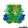


 PDBj
PDBj
 Assembly


 Mass: 39.098 Da / Num. of mol.: 5 / Source method: obtained synthetically / Formula: K
 Mass: 24.305 Da / Num. of mol.: 1 / Source method: obtained synthetically / Formula: Mg
 Mass: 40.078 Da / Num. of mol.: 2 / Source method: obtained synthetically / Formula: Ca
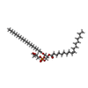
 Mass: 749.007 Da / Num. of mol.: 15 / Source method: obtained synthetically / Formula: C40H77O10P / Comment: phospholipid*YM
 Sample preparation
 Electron microscopy imaging
 Processing