 Movie
Movie Controller

[English] 日本語
 Yorodumi
- PDB-5tc1: In situ structures of the genome and genome-delivery apparatus in... -
+ Open data

- Basic information
| Entry | Database: PDB / ID: 5tc1 | ||||||
|---|---|---|---|---|---|---|---|
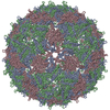
| Title | In situ structures of the genome and genome-delivery apparatus in ssRNA bacteriophage MS2 | ||||||
 Components Components |
| ||||||
 Keywords Keywords |  viral protein/rna / asymmetric cryoEM reconstruction / ssRNA genome structure / genome-delivery apparatus / genome-capsid interactions / viral protein-rna complex viral protein/rna / asymmetric cryoEM reconstruction / ssRNA genome structure / genome-delivery apparatus / genome-capsid interactions / viral protein-rna complex | ||||||
| Function / homology |  Function and homology information Function and homology informationviral genome circularization / virion attachment to host cell pilus / negative regulation of viral translation / T=3 icosahedral viral capsid / virion component / regulation of translation / symbiont entry into host cell / structural molecule activity / RNA binding / identical protein bindingSimilarity search - Function | ||||||
| Biological species |  Enterobacteria phage MS2 (virus) Enterobacteria phage MS2 (virus) | ||||||
| Method | ELECTRON MICROSCOPY / single particle reconstruction / cryo EM / Resolution: 3.6 Å | ||||||
 Authors Authors | Dai, X.H. / Li, Z.H. / Lai, M. / Shu, S. / Du, Y.S. / Zhou, Z.H. / Sun, R. | ||||||
 Citation Citation | Journal: Nature / Year: 2017 Title: In situ structures of the genome and genome-delivery apparatus in a single-stranded RNA virus. Authors: Xinghong Dai / Zhihai Li / Mason Lai / Sara Shu / Yushen Du / Z Hong Zhou / Ren Sun /   Abstract: Packaging of the genome into a protein capsid and its subsequent delivery into a host cell are two fundamental processes in the life cycle of a virus. Unlike double-stranded DNA viruses, which pump ...Packaging of the genome into a protein capsid and its subsequent delivery into a host cell are two fundamental processes in the life cycle of a virus. Unlike double-stranded DNA viruses, which pump their genome into a preformed capsid, single-stranded RNA (ssRNA) viruses, such as bacteriophage MS2, co-assemble their capsid with the genome; however, the structural basis of this co-assembly is poorly understood. MS2 infects Escherichia coli via the host 'sex pilus' (F-pilus); it was the first fully sequenced organism and is a model system for studies of translational gene regulation, RNA-protein interactions, and RNA virus assembly. Its positive-sense ssRNA genome of 3,569 bases is enclosed in a capsid with one maturation protein monomer and 89 coat protein dimers arranged in a T = 3 icosahedral lattice. The maturation protein is responsible for attaching the virus to an F-pilus and delivering the viral genome into the host during infection, but how the genome is organized and delivered is not known. Here we describe the MS2 structure at 3.6 Å resolution, determined by electron-counting cryo-electron microscopy (cryoEM) and asymmetric reconstruction. We traced approximately 80% of the backbone of the viral genome, built atomic models for 16 RNA stem-loops, and identified three conserved motifs of RNA-coat protein interactions among 15 of these stem-loops with diverse sequences. The stem-loop at the 3' end of the genome interacts extensively with the maturation protein, which, with just a six-helix bundle and a six-stranded β-sheet, forms a genome-delivery apparatus and joins 89 coat protein dimers to form a capsid. This atomic description of genome-capsid interactions in a spherical ssRNA virus provides insight into genome delivery via the host sex pilus and mechanisms underlying ssRNA-capsid co-assembly, and inspires speculation about the links between nucleoprotein complexes and the origins of viruses. | ||||||
| History |
|
- Structure visualization
| Movie |
Movie viewer |
|---|---|
| Structure viewer | Molecule: MolmilJmol/JSmol |
- Downloads & links
-Download
| PDBx/mmCIF format | 5tc1.cif.gz | 411.3 KB | Display | PDBx/mmCIF format |
|---|---|---|---|---|
| PDB format | pdb5tc1.ent.gz | 304.8 KB | Display | PDB format |
| PDBx/mmJSON format | 5tc1.json.gz | Tree view | PDBx/mmJSON format | |
| Others |  Other downloads Other downloads |
-Validation report
| Arichive directory | https://data.pdbj.org/pub/pdb/validation_reports/tc/5tc1ftp://data.pdbj.org/pub/pdb/validation_reports/tc/5tc1 | HTTPS FTP |
|---|
-Related structure data
| Related structure data | 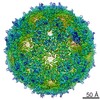 8397MC M: map data used to model this data C: citing same article ( |
|---|---|
| Similar structure data |
-Links
 PDBj
PDBj


- Assembly
| Deposited unit | 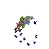
|
|---|---|
| 1 |
|
| Details | The MS2 virion contains an ssRNA chain of the genome enclosed by a capsid built from 178 copies of coat protein and a monomer of the maturation protein. Our structure shows that the maturation protein breaks the icosahedral symmetry of the capsid and imparts structural changes to some of its neighboring coat proteins (chains D, E, F, G, H in this model). All other 173 copies of the coat protein conform to the icosahedral symmetry and have same structure with the crystallographic structure of MS2 capsid with PDB ID 2MS2. Only three of these 173 chains are included in the model (chains A, B, C) for reference. |
-Components
| #1: Protein | Capsid / Coat protein Mass: 13869.659 Da / Num. of mol.: 8 / Source method: isolated from a natural source Details: The capsid of MS2 contains 178 copies of the coat protein arranged as 89 dimers in a T=3 icosahedral lattice. Structure of the capsid has been solved by crystallography with icosahedral ...Details: The capsid of MS2 contains 178 copies of the coat protein arranged as 89 dimers in a T=3 icosahedral lattice. Structure of the capsid has been solved by crystallography with icosahedral symmetry applied (PDB ID 2MS2, also included as chains A, B, C in this model). Chains D, E, F, G, H are coat proteins that have slightly different structures in the asymmetric cryoEM reconstruction compared to the crystallographic structure. Structures of the other 170 copies of the coat protein are the same with 2MS2, and thus are not included due to the limited number of chain IDs. Source: (natural) Enterobacteria phage MS2 (virus) / References: UniProt: P03612#2: Protein | | Mass: 44030.934 Da / Num. of mol.: 1 / Source method: isolated from a natural source Details: The capsid of MS2 contains a single copy of the maturation protein. Our structure shows that it replaces a coat protein dimer at one of the 2-fold icosahedral symmetry axes. Function of the ...Details: The capsid of MS2 contains a single copy of the maturation protein. Our structure shows that it replaces a coat protein dimer at one of the 2-fold icosahedral symmetry axes. Function of the maturation protein is to attach the MS2 virion to the host (E. coli) F-pili and deliver the ssRNA viral genome into the host during infection. of the icosahedral capsid. 178 copies of the coat protein arranged as 89 dimers in a T=3 icosahedral lattice. Structure of the capsid has been solved by crystallography with icosahedral symmetry applied (PDB ID 2MS2, also included as chains A, B, C in this model). Chains D, E, F, G, H are coat proteins that have slightly different structures in the asymmetric cryoEM reconstruction compared to the crystallographic structure. Structures of the other 170 copies of the coat protein are the same with 2MS2, and thus are not included due to the limited number of chain IDs. Source: (natural) Enterobacteria phage MS2 (virus) / References: UniProt: P03610#3: RNA chain | | Mass: 1147200.750 Da / Num. of mol.: 1 / Source method: isolated from a natural source / Source: (natural) Enterobacteria phage MS2 (virus) / References: GenBank: 15081 |
|---|
-Experimental details
-Experiment
| Experiment | Method: ELECTRON MICROSCOPY |
|---|---|
| EM experiment | Aggregation state: PARTICLE / 3D reconstruction method: single particle reconstruction |
- Sample preparation
| Component |
| ||||||||||||||||||||||||||||||||||||||||||
|---|---|---|---|---|---|---|---|---|---|---|---|---|---|---|---|---|---|---|---|---|---|---|---|---|---|---|---|---|---|---|---|---|---|---|---|---|---|---|---|---|---|---|---|
| Molecular weight |
| ||||||||||||||||||||||||||||||||||||||||||
| Source (natural) |
| ||||||||||||||||||||||||||||||||||||||||||
| Details of virus |
| ||||||||||||||||||||||||||||||||||||||||||
| Natural host |
| ||||||||||||||||||||||||||||||||||||||||||
| Virus shell |
| ||||||||||||||||||||||||||||||||||||||||||
| Buffer solution | pH: 7.4 / Details: pH7.4 PBS | ||||||||||||||||||||||||||||||||||||||||||
| Specimen | Embedding applied: NO / Shadowing applied: NO / Staining applied: NO / Vitrification applied: YES | ||||||||||||||||||||||||||||||||||||||||||
| Specimen support | Grid material: COPPER / Grid mesh size: 300 divisions/in. / Grid type: Quantifoil R1.2/1.3 | ||||||||||||||||||||||||||||||||||||||||||
| Vitrification | Instrument: FEI VITROBOT MARK II / Cryogen name: ETHANE / Humidity: 100 % / Chamber temperature: 295 K |
- Electron microscopy imaging
| Experimental equipment |  Model: Titan Krios / Image courtesy: FEI Company |
|---|---|
| Microscopy | Model: FEI TITAN KRIOS / Details: EFTEM mode with Gatan GIF energy filter. |
| Electron gun | Electron source: FIELD EMISSION GUN / Accelerating voltage: 300 kV / Illumination mode: FLOOD BEAM |
| Electron lens | Mode: BRIGHT FIELDBright-field microscopy / Nominal magnification: 130000 X / Calibrated magnification: 47170 X / Cs: 2.7 mm / C2 aperture diameter: 50 µm / Alignment procedure: BASIC |
| Specimen holder | Cryogen: NITROGEN / Specimen holder model: FEI TITAN KRIOS AUTOGRID HOLDER / Temperature (min): 79 K |
| Image recording | Average exposure time: 5.8 sec. / Electron dose: 50 e/Å2 / Detector mode: SUPER-RESOLUTION / Film or detector model: GATAN K2 SUMMIT (4k x 4k) / Num. of grids imaged: 2 / Num. of real images: 6080 |
| EM imaging optics | Energyfilter name: GIF / Energyfilter upper: 20 eV / Energyfilter lower: 20 eV |
| Image scans | Sampling size: 5 µm / Width: 7676 / Height: 7420 / Movie frames/image: 29 / Used frames/image: 1-14 |
- Processing
| EM software |
| |||||||||||||||||||||||||||||||||||||||||||||
|---|---|---|---|---|---|---|---|---|---|---|---|---|---|---|---|---|---|---|---|---|---|---|---|---|---|---|---|---|---|---|---|---|---|---|---|---|---|---|---|---|---|---|---|---|---|---|
| CTF correction | Type: PHASE FLIPPING AND AMPLITUDE CORRECTION | |||||||||||||||||||||||||||||||||||||||||||||
| Particle selection | Num. of particles selected: 360000 | |||||||||||||||||||||||||||||||||||||||||||||
| Symmetry | Point symmetry: C1 (asymmetric) | |||||||||||||||||||||||||||||||||||||||||||||
| 3D reconstruction | Resolution: 3.6 Å / Resolution method: FSC 0.143 CUT-OFF / Num. of particles: 339718 / Algorithm: FOURIER SPACE / Num. of class averages: 1 / Symmetry type: POINT | |||||||||||||||||||||||||||||||||||||||||||||
| Atomic model building | Protocol: AB INITIO MODEL / Space: REAL |
