 Movie
Movie Controller


+ Open data

- Basic information
| Entry | Database: PDB / ID: 5oq0 | ||||||
|---|---|---|---|---|---|---|---|
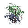
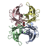
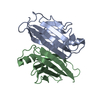
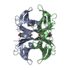
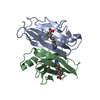
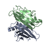
| Title | Crystal structure of transthyretin mutant 87-110-117 | ||||||
 Components Components | Transthyretin | ||||||
 Keywords Keywords | TRANSPORT PROTEIN / Amyloidosis / homo-tetramer / retinol-binding protein / TTR | ||||||
| Function / homology |  Function and homology information Function and homology informationRetinoid cycle disease events / thyroid hormone binding / The canonical retinoid cycle in rods (twilight vision) / Non-integrin membrane-ECM interactions / purine nucleobase metabolic process / Retinoid metabolism and transport / hormone activity / azurophil granule lumen / Amyloid fiber formation / Neutrophil degranulation ...Retinoid cycle disease events / thyroid hormone binding / The canonical retinoid cycle in rods (twilight vision) / Non-integrin membrane-ECM interactions / purine nucleobase metabolic process / Retinoid metabolism and transport / hormone activity / azurophil granule lumen / Amyloid fiber formation / Neutrophil degranulation / extracellular space / extracellular exosome / extracellular region / identical protein bindingSimilarity search - Function | ||||||
| Biological species |  Homo sapiens (human) Homo sapiens (human) | ||||||
| Method | X-RAY DIFFRACTION / SYNCHROTRON / MOLECULAR REPLACEMENT / Resolution: 1.94 Å | ||||||
 Authors Authors | Zanotti, G. / Vallese, F. / Berni, R. | ||||||
 Citation Citation | Journal: PLoS ONE / Year: 2017 Title: Structural and dynamics evidence for scaffold asymmetric flexibility of the human transthyretin tetramer. Authors: Zanotti, G. / Vallese, F. / Ferrari, A. / Menozzi, I. / Saldano, T.E. / Berto, P. / Fernandez-Alberti, S. / Berni, R. | ||||||
| History |
|
- Structure visualization
| Structure viewer | Molecule: MolmilJmol/JSmol |
|---|
- Downloads & links
-Download
| PDBx/mmCIF format | 5oq0.cif.gz | 59.7 KB | Display | PDBx/mmCIF format |
|---|---|---|---|---|
| PDB format | pdb5oq0.ent.gz | 43.1 KB | Display | PDB format |
| PDBx/mmJSON format | 5oq0.json.gz | Tree view | PDBx/mmJSON format | |
| Others |  Other downloads Other downloads |
-Validation report
| Arichive directory | https://data.pdbj.org/pub/pdb/validation_reports/oq/5oq0ftp://data.pdbj.org/pub/pdb/validation_reports/oq/5oq0 | HTTPS FTP |
|---|
-Related structure data
| Related structure data |  4wo0S S: Starting model for refinement |
|---|---|
| Similar structure data |


-Links
 PDBj
PDBj


- Assembly
| Deposited unit | 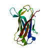
| ||||||||
|---|---|---|---|---|---|---|---|---|---|
| 1 | 
| ||||||||
| Unit cell |
| ||||||||
| Components on special symmetry positions |
|
-Components
| #1: Protein | / ATTR / Prealbumin / TBPA Mass: 13821.456 Da / Num. of mol.: 1 / Mutation: F87M/L110M/S117E Source method: isolated from a genetically manipulated source Source: (gene. exp.) Homo sapiens (human) / Gene: TTR, PALB / Production host:  Escherichia coli BL21(DE3) (bacteria) / References: UniProt: P02766 Escherichia coli BL21(DE3) (bacteria) / References: UniProt: P02766 |
|---|---|
| #2: Water | ChemComp-HOH / Water Mass: 18.015 Da / Num. of mol.: 27 / Source method: isolated from a natural source / Formula: H2O Mass: 18.015 Da / Num. of mol.: 27 / Source method: isolated from a natural source / Formula: H2O |
-Experimental details
-Experiment
| Experiment | Method: X-RAY DIFFRACTION / Number of used crystals: 1 |
|---|
- Sample preparation
| Crystal | Density Matthews: 2.14 Å3/Da / Density % sol: 42.54 % |
|---|---|
| Crystal grow | Temperature: 293 K / Method: vapor diffusion, hanging drop Details: 0.1 M sodium phosphate (pH 7.5), 2.2 M ammonium sulphate PH range: 7.5 |
-Data collection
| Diffraction | Mean temperature: 100 K |
|---|---|
| Diffraction source | Source: SYNCHROTRON / Site: ESRF  / Beamline: ID30B / Wavelength: 0.973186 Å / Beamline: ID30B / Wavelength: 0.973186 Å |
| Detector | Type: DECTRIS PILATUS3 S 6M / Detector: PIXEL / Date: Aug 10, 2016 |
| Radiation | Protocol: SINGLE WAVELENGTH / Monochromatic (M) / Laue (L): M / Scattering type: x-ray |
| Radiation wavelength | Wavelength: 0.973186 Å / Relative weight: 1 |
| Reflection | Resolution: 1.94→52.29 Å / Num. obs: 9125 / % possible obs: 97.4 % / Observed criterion σ(F): 0 / Observed criterion σ(I): 0 / Redundancy: 7.2 % / Rmerge(I) obs: 0.073 / Rpim(I) all: 0.03 / Net I/σ(I): 13 |
| Reflection shell | Resolution: 1.94→2.01 Å / Redundancy: 4.8 % / Rmerge(I) obs: 0.916 / Mean I/σ(I) obs: 1.6 / Rpim(I) all: 0.514 / % possible all: 80.5 |
- Processing
| Software |
| ||||||||||||||||||||||||||||||||||||||||
|---|---|---|---|---|---|---|---|---|---|---|---|---|---|---|---|---|---|---|---|---|---|---|---|---|---|---|---|---|---|---|---|---|---|---|---|---|---|---|---|---|---|
| Refinement | Method to determine structure: MOLECULAR REPLACEMENT Starting model: 4WO0 Resolution: 1.94→41.783 Å / SU ML: 0.28 / Cross valid method: FREE R-VALUE / σ(F): 1.35 / Phase error: 37.57
| ||||||||||||||||||||||||||||||||||||||||
| Solvent computation | Shrinkage radii: 0.9 Å / VDW probe radii: 1.11 Å | ||||||||||||||||||||||||||||||||||||||||
| Refinement step | Cycle: LAST / Resolution: 1.94→41.783 Å
| ||||||||||||||||||||||||||||||||||||||||
| Refine LS restraints |
| ||||||||||||||||||||||||||||||||||||||||
| LS refinement shell |
| ||||||||||||||||||||||||||||||||||||||||
| Refinement TLS params. | Method: refined / Origin x: 43.0719 Å / Origin y: 90.1711 Å / Origin z: 12.8608 Å
| ||||||||||||||||||||||||||||||||||||||||
| Refinement TLS group | Selection details: all |
