- PDB-5ome: The cryofrozen atomic resolution X-ray crystal structure of the r... -
+
Open data
ID or keywords:
Loading...
-
Basic information
Entry
Database: PDB / ID: 5ome
Title
The cryofrozen atomic resolution X-ray crystal structure of the reduced form (Fe2+) perdeuterated Pyrococcus furiosus Rubredoxin in D2O (100K, 0.75 Angstrom resolution)
Components
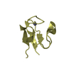
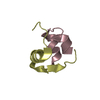
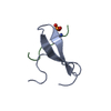
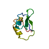
Rubredoxin
Keywords
ELECTRON TRANSPORT / Perdeuterated rubredoxin / pyrococcus furiosus / atomic resolution / cryofrozen / reduced / iron
Mass: 18.015 Da / Num. of mol.: 209 / Source method: isolated from a natural source / Formula: H2O
-
Experimental details
-
Experiment
Experiment
Method: X-RAY DIFFRACTION / Number of used crystals: 1
-
Sample preparation
Crystal
Density Matthews: 2.12 Å3/Da / Density % sol: 41.98 % Description: bipyramid bright red (Fe3+), Subsequently reduced with Na2S2O3 to get transparent (Fe2+) crystal. X-ray data collection performed with helicoidal strategy over 180 degrees rotation.
Method to determine structure: AB INITIO PHASING / Resolution: 0.747→18.363 Å / SU ML: 0.02 / Cross valid method: FREE R-VALUE / σ(F): 1.35 / Phase error: 10.12 Details: Deuterium atoms are isotropic, protein atoms, Fe atom and solvent O atoms are anisotropic
Rfactor
Num. reflection
% reflection
Selection details
Rfree
0.1214
2892
5.03 %
random
Rwork
0.111
-
-
-
obs
0.1115
57545
85.72 %
-
Solvent computation
Shrinkage radii: 0.9 Å / VDW probe radii: 1.11 Å
Refinement step
Cycle: LAST / Resolution: 0.747→18.363 Å
Protein
Nucleic acid
Ligand
Solvent
Total
Num. atoms
421
0
12
209
642
Refine LS restraints
Refine-ID
Type
Dev ideal
Number
X-RAY DIFFRACTION
f_bond_d
0.008
498
X-RAY DIFFRACTION
f_angle_d
1.01
687
X-RAY DIFFRACTION
f_dihedral_angle_d
17.92
191
X-RAY DIFFRACTION
f_chiral_restr
0.084
64
X-RAY DIFFRACTION
f_plane_restr
0.009
96
LS refinement shell
Resolution (Å)
Rfactor Rfree
Num. reflection Rfree
Rfactor Rwork
Num. reflection Rwork
Refine-ID
% reflection obs (%)
0.747-0.7593
0.2897
15
0.1935
261
X-RAY DIFFRACTION
9
0.7593-0.7723
0.14
50
0.1481
865
X-RAY DIFFRACTION
29
0.7723-0.7864
0.143
59
0.1361
1360
X-RAY DIFFRACTION
45
0.7864-0.8015
0.1257
83
0.125
1848
X-RAY DIFFRACTION
61
0.8015-0.8179
0.1252
142
0.1124
2226
X-RAY DIFFRACTION
75
0.8179-0.8357
0.1046
130
0.111
2640
X-RAY DIFFRACTION
87
0.8357-0.8551
0.1109
166
0.1058
2831
X-RAY DIFFRACTION
95
0.8551-0.8765
0.1075
135
0.1008
3003
X-RAY DIFFRACTION
100
0.8765-0.9002
0.1136
153
0.0936
3025
X-RAY DIFFRACTION
100
0.9002-0.9267
0.102
162
0.0914
2991
X-RAY DIFFRACTION
100
0.9267-0.9566
0.102
153
0.0899
3027
X-RAY DIFFRACTION
100
0.9566-0.9908
0.1112
171
0.0856
3013
X-RAY DIFFRACTION
100
0.9908-1.0304
0.0969
151
0.087
3034
X-RAY DIFFRACTION
100
1.0304-1.0773
0.0942
176
0.0853
2994
X-RAY DIFFRACTION
100
1.0773-1.1341
0.0926
149
0.0875
3048
X-RAY DIFFRACTION
100
1.1341-1.2052
0.1019
176
0.0936
3017
X-RAY DIFFRACTION
100
1.2052-1.2982
0.1048
184
0.1041
3022
X-RAY DIFFRACTION
100
1.2982-1.4288
0.1148
161
0.1164
3053
X-RAY DIFFRACTION
100
1.4288-1.6354
0.1324
146
0.1208
3097
X-RAY DIFFRACTION
100
1.6354-2.06
0.1291
168
0.13
3096
X-RAY DIFFRACTION
99
2.06-18.3683
0.1543
162
0.126
3202
X-RAY DIFFRACTION
98
+
About Yorodumi
-
News
-
Feb 9, 2022. New format data for meta-information of EMDB entries
New format data for meta-information of EMDB entries
Version 3 of the EMDB header file is now the official format.
The previous official version 1.9 will be removed from the archive.
In the structure databanks used in Yorodumi, some data are registered as the other names, "COVID-19 virus" and "2019-nCoV". Here are the details of the virus and the list of structure data.
Jan 31, 2019. EMDB accession codes are about to change! (news from PDBe EMDB page)
EMDB accession codes are about to change! (news from PDBe EMDB page)
The allocation of 4 digits for EMDB accession codes will soon come to an end. Whilst these codes will remain in use, new EMDB accession codes will include an additional digit and will expand incrementally as the available range of codes is exhausted. The current 4-digit format prefixed with “EMD-” (i.e. EMD-XXXX) will advance to a 5-digit format (i.e. EMD-XXXXX), and so on. It is currently estimated that the 4-digit codes will be depleted around Spring 2019, at which point the 5-digit format will come into force.
The EM Navigator/Yorodumi systems omit the EMD- prefix.
Related info.:Q: What is EMD? / ID/Accession-code notation in Yorodumi/EM Navigator
Yorodumi is a browser for structure data from EMDB, PDB, SASBDB, etc.
This page is also the successor to EM Navigator detail page, and also detail information page/front-end page for Omokage search.
The word "yorodu" (or yorozu) is an old Japanese word meaning "ten thousand". "mi" (miru) is to see.
Related info.:EMDB / PDB / SASBDB / Comparison of 3 databanks / Yorodumi Search / Aug 31, 2016. New EM Navigator & Yorodumi / Yorodumi Papers / Jmol/JSmol / Function and homology information / Changes in new EM Navigator and Yorodumi
 Movie
Movie Controller

 Yorodumi
 Open data
 Basic information
 Components
 Keywords
 Function and homology information

 Authors
 United Kingdom,
  France, 4items
  Citation
 Structure visualization
 Downloads & links
 Other downloads


 PDBj
PDBj
 Assembly


 Mass: 55.845 Da / Num. of mol.: 1 / Source method: obtained synthetically / Formula: Fe
 Mass: 22.990 Da / Num. of mol.: 1 / Source method: obtained synthetically / Formula: Na
 Mass: 94.971 Da / Num. of mol.: 2 / Source method: obtained synthetically / Formula: PO4
 Mass: 18.015 Da / Num. of mol.: 209 / Source method: isolated from a natural source / Formula: H2O
 Sample preparation
 Processing