[English] 日本語
Yorodumi
- PDB-5o9x: Crystal structure of Aspergillus fumigatus N-acetylphosphoglucosa... -

+
Open data


ID or keywords:

Loading...

-
Basic information

Entry
Database: PDB / ID: 5o9x
TitleCrystal structure of Aspergillus fumigatus N-acetylphosphoglucosamine mutate S69A in complex with glucose1,6bisphosphate
ComponentsPhosphoacetylglucosamine mutase
KeywordsTRANSFERASE / Mutase / N-acetylphosphoglucosamine / Aspergillus fumigatus
Function / homology
Function and homology information


phosphoacetylglucosamine mutase / phosphoacetylglucosamine mutase activity / UDP-N-acetylglucosamine biosynthetic process / carbohydrate metabolic process / magnesium ion binding
Similarity search - Function
Phosphoacetylglucosamine mutase / Alpha-D-phosphohexomutase, C-terminal / Phosphoglucomutase/phosphomannomutase, C-terminal domain / Alpha-D-Glucose-1,6-Bisphosphate; Chain A, domain 3 / Alpha-D-Glucose-1,6-Bisphosphate, subunit A, domain 3 / Alpha-D-phosphohexomutase, C-terminal domain / Alpha-D-phosphohexomutase, conserved site / Phosphoglucomutase and phosphomannomutase phosphoserine signature. / Alpha-D-phosphohexomutase, alpha/beta/alpha domain I / Alpha-D-phosphohexomutase, alpha/beta/alpha I/II/III ...Phosphoacetylglucosamine mutase / Alpha-D-phosphohexomutase, C-terminal / Phosphoglucomutase/phosphomannomutase, C-terminal domain / Alpha-D-Glucose-1,6-Bisphosphate; Chain A, domain 3 / Alpha-D-Glucose-1,6-Bisphosphate, subunit A, domain 3 / Alpha-D-phosphohexomutase, C-terminal domain / Alpha-D-phosphohexomutase, conserved site / Phosphoglucomutase and phosphomannomutase phosphoserine signature. / Alpha-D-phosphohexomutase, alpha/beta/alpha domain I / Alpha-D-phosphohexomutase, alpha/beta/alpha I/II/III / Alpha-D-phosphohexomutase, C-terminal domain superfamily / Phosphoglucomutase/phosphomannomutase, alpha/beta/alpha domain I / TATA-Binding Protein / 2-Layer Sandwich / 3-Layer(aba) Sandwich / Alpha Beta
Similarity search - Domain/homology
1,6-di-O-phosphono-alpha-D-glucopyranose / Phosphoacetylglucosamine mutase
Similarity search - Component
Biological speciesAspergillus lentulus (mold)
MethodX-RAY DIFFRACTION / SYNCHROTRON / Resolution: 1.9 Å
AuthorsRaimi, O.G. / Hurtado-Guerrero, R.
CitationJournal: Biochem. J. / Year: 2018
Title: Evidence for substrate-assisted catalysis inN-acetylphosphoglucosamine mutase.
Authors: Raimi, O.G. / Hurtado-Guerrero, R. / van Aalten, D.M.F.
History
DepositionJun 20, 2017Deposition site: PDBE / Processing site: PDBE
Revision 1.0Jul 11, 2018Provider: repository / Type: Initial release
Revision 1.1Aug 29, 2018Group: Data collection / Database references / Category: citation / citation_author
Item: _citation.journal_volume / _citation.page_first ..._citation.journal_volume / _citation.page_first / _citation.page_last / _citation.title / _citation_author.name
Revision 1.2Jul 29, 2020Group: Data collection / Derived calculations / Structure summary
Category: chem_comp / entity ...chem_comp / entity / pdbx_chem_comp_identifier / pdbx_entity_nonpoly / struct_site / struct_site_gen
Item: _chem_comp.mon_nstd_flag / _chem_comp.name ..._chem_comp.mon_nstd_flag / _chem_comp.name / _entity.pdbx_description / _pdbx_entity_nonpoly.name
Description: Carbohydrate remediation / Provider: repository / Type: Remediation

-
Structure visualization

Structure viewerMolecule:
MolmilJmol/JSmol

Downloads & links

-
Assembly

Deposited unit
A: Phosphoacetylglucosamine mutase
hetero molecules


Theoretical massNumber of molelcules
Total (without water)60,5076
Polymers59,9131
Non-polymers5945
Water8,485471
1


  • Idetical with deposited unit
  • defined by author&software
  • Evidence: gel filtration
TypeNameSymmetry operationNumber
identity operation1_555x,y,z1
Buried area1380 Å2
ΔGint-28 kcal/mol
Surface area21720 Å2
MethodPISA
Unit cell
Length a, b, c (Å)80.860, 87.574, 91.867
Angle α, β, γ (deg.)90.00, 90.00, 90.00
Int Tables number19
Space group name H-MP212121

-
Components

#1: Protein Phosphoacetylglucosamine mutase / / PAGM / Acetylglucosamine phosphomutase / N-acetylglucosamine-phosphate mutase


Mass: 59912.953 Da / Num. of mol.: 1 / Mutation: S69A
Source method: isolated from a genetically manipulated source
Source: (gene. exp.) Aspergillus lentulus (mold) / Gene: ALT_8497 / Plasmid: pGEX-6P / Production host: Escherichia coli BL21(DE3) (bacteria) / Variant (production host): PlysS
References: UniProt: A0A0S7E9S6, phosphoacetylglucosamine mutase
#2: Sugar ChemComp-G16 / 1,6-di-O-phosphono-alpha-D-glucopyranose / Glucose 1,6-bisphosphate


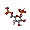
Type: D-saccharide / Mass: 339.108 Da / Num. of mol.: 1
Source method: isolated from a genetically manipulated source
Formula: C6H13O12P2
IdentifierTypeProgram
a-D-Glcp1PO36PO3IUPAC CARBOHYDRATE SYMBOLPDB-CARE 1.0
#3: Chemical ChemComp-CL / CHLORIDE ION / Chloride


Mass: 35.453 Da / Num. of mol.: 2 / Source method: obtained synthetically / Formula: Cl
#4: Chemical ChemComp-GOL / GLYCEROL / GLYCERIN / PROPANE-1,2,3-TRIOL / Glycerol


Mass: 92.094 Da / Num. of mol.: 2 / Source method: obtained synthetically / Formula: C3H8O3
#5: Water ChemComp-HOH / water / Water


Mass: 18.015 Da / Num. of mol.: 471 / Source method: isolated from a natural source / Formula: H2O

-
Experimental details

-
Experiment

ExperimentMethod: X-RAY DIFFRACTION / Number of used crystals: 1

-
Sample preparation

CrystalDensity Matthews: 2.71 Å3/Da / Density % sol: 54.68 %
Crystal growTemperature: 291 K / Method: vapor diffusion, sitting drop
Details: 1. 0.2 M sodium thiocyanate, 20% PEG 3350 and 0.1 M sodium bromide 2. 0.10 M glycine, 22% PEG 3350 and 0.2 M sodium thiocyanate

-
Data collection

DiffractionMean temperature: 100 K
Diffraction sourceSource: SYNCHROTRON / Site: ESRF / Beamline: ID14-2 / Wavelength: 0.933 Å
DetectorType: ADSC QUANTUM 4 / Detector: CCD / Date: Apr 11, 2007
RadiationProtocol: SINGLE WAVELENGTH / Monochromatic (M) / Laue (L): M / Scattering type: x-ray
Radiation wavelengthWavelength: 0.933 Å / Relative weight: 1
ReflectionResolution: 1.9→20 Å / Num. obs: 51704 / % possible obs: 97.6 % / Redundancy: 3.7 % / Net I/σ(I): 19.2
Reflection shellResolution: 1.9→1.97 Å / Redundancy: 3.6 % / Num. unique obs: 4939 / % possible all: 94.4

-
Processing

Software
NameVersionClassification
REFMAC5.8.0158refinement
XDSdata reduction
SCALAdata scaling
MOLREPphasing
RefinementResolution: 1.9→20 Å / Cor.coef. Fo:Fc: 0.963 / Cor.coef. Fo:Fc free: 0.947 / SU B: 2.997 / SU ML: 0.087 / Cross valid method: THROUGHOUT / ESU R: 0.122 / ESU R Free: 0.118 / Stereochemistry target values: MAXIMUM LIKELIHOOD / Details: HYDROGENS HAVE BEEN USED IF PRESENT IN THE INPUT
RfactorNum. reflection% reflectionSelection details
Rfree0.20123 519 1 %RANDOM
Rwork0.1649 ---
obs0.1653 50268 97.45 %-
Solvent computationIon probe radii: 0.8 Å / Shrinkage radii: 0.8 Å / VDW probe radii: 1.4 Å / Solvent model: MASK
Displacement parametersBiso mean: 26.934 Å2
Baniso -1Baniso -2Baniso -3
1-1.21 Å20 Å20 Å2
2---0.03 Å20 Å2
3----1.18 Å2
Refinement stepCycle: 1 / Resolution: 1.9→20 Å
ProteinNucleic acidLigandSolventTotal
Num. atoms4144 0 34 471 4649
Refine LS restraints
Refine-IDTypeDev idealDev ideal targetNumber
X-RAY DIFFRACTIONr_bond_refined_d0.0230.0194253
X-RAY DIFFRACTIONr_bond_other_d
X-RAY DIFFRACTIONr_angle_refined_deg2.1241.9775762
X-RAY DIFFRACTIONr_angle_other_deg
X-RAY DIFFRACTIONr_dihedral_angle_1_deg6.4295542
X-RAY DIFFRACTIONr_dihedral_angle_2_deg33.29624.076184
X-RAY DIFFRACTIONr_dihedral_angle_3_deg15.53215727
X-RAY DIFFRACTIONr_dihedral_angle_4_deg17.5291531
X-RAY DIFFRACTIONr_chiral_restr0.160.2663
X-RAY DIFFRACTIONr_gen_planes_refined0.0110.0213174
X-RAY DIFFRACTIONr_gen_planes_other
X-RAY DIFFRACTIONr_nbd_refined
X-RAY DIFFRACTIONr_nbd_other
X-RAY DIFFRACTIONr_nbtor_refined
X-RAY DIFFRACTIONr_nbtor_other
X-RAY DIFFRACTIONr_xyhbond_nbd_refined
X-RAY DIFFRACTIONr_xyhbond_nbd_other
X-RAY DIFFRACTIONr_metal_ion_refined
X-RAY DIFFRACTIONr_metal_ion_other
X-RAY DIFFRACTIONr_symmetry_vdw_refined
X-RAY DIFFRACTIONr_symmetry_vdw_other
X-RAY DIFFRACTIONr_symmetry_hbond_refined
X-RAY DIFFRACTIONr_symmetry_hbond_other
X-RAY DIFFRACTIONr_symmetry_metal_ion_refined
X-RAY DIFFRACTIONr_symmetry_metal_ion_other
X-RAY DIFFRACTIONr_mcbond_it2.482.3982165
X-RAY DIFFRACTIONr_mcbond_other
X-RAY DIFFRACTIONr_mcangle_it3.3923.5792705
X-RAY DIFFRACTIONr_mcangle_other
X-RAY DIFFRACTIONr_scbond_it4.1812.8082088
X-RAY DIFFRACTIONr_scbond_other
X-RAY DIFFRACTIONr_scangle_it
X-RAY DIFFRACTIONr_scangle_other
X-RAY DIFFRACTIONr_long_range_B_refined6.98434.2486786
X-RAY DIFFRACTIONr_long_range_B_other
X-RAY DIFFRACTIONr_rigid_bond_restr
X-RAY DIFFRACTIONr_sphericity_free
X-RAY DIFFRACTIONr_sphericity_bonded
LS refinement shellResolution: 1.9→1.949 Å / Total num. of bins used: 20
RfactorNum. reflection% reflection
Rfree0.265 27 -
Rwork0.218 3529 -
obs--94.2 %

+
About Yorodumi

-
News

-
Feb 9, 2022. New format data for meta-information of EMDB entries

New format data for meta-information of EMDB entries

  • Version 3 of the EMDB header file is now the official format.
  • The previous official version 1.9 will be removed from the archive.

Related info.:EMDB header

External links:wwPDB to switch to version 3 of the EMDB data model

-
Aug 12, 2020. Covid-19 info

Covid-19 info

URL: https://pdbj.org/emnavi/covid19.php

New page: Covid-19 featured information page in EM Navigator.

Related info.:Covid-19 info / Mar 5, 2020. Novel coronavirus structure data

+
Mar 5, 2020. Novel coronavirus structure data

Novel coronavirus structure data

Related info.:Yorodumi Speices / Aug 12, 2020. Covid-19 info

External links:COVID-19 featured content - PDBj / Molecule of the Month (242):Coronavirus Proteases

+
Jan 31, 2019. EMDB accession codes are about to change! (news from PDBe EMDB page)

EMDB accession codes are about to change! (news from PDBe EMDB page)

  • The allocation of 4 digits for EMDB accession codes will soon come to an end. Whilst these codes will remain in use, new EMDB accession codes will include an additional digit and will expand incrementally as the available range of codes is exhausted. The current 4-digit format prefixed with “EMD-” (i.e. EMD-XXXX) will advance to a 5-digit format (i.e. EMD-XXXXX), and so on. It is currently estimated that the 4-digit codes will be depleted around Spring 2019, at which point the 5-digit format will come into force.
  • The EM Navigator/Yorodumi systems omit the EMD- prefix.

Related info.:Q: What is EMD? / ID/Accession-code notation in Yorodumi/EM Navigator

External links:EMDB Accession Codes are Changing Soon! / Contact to PDBj

+
Jul 12, 2017. Major update of PDB

Major update of PDB

  • wwPDB released updated PDB data conforming to the new PDBx/mmCIF dictionary.
  • This is a major update changing the version number from 4 to 5, and with Remediation, in which all the entries are updated.
  • In this update, many items about electron microscopy experimental information are reorganized (e.g. em_software).
  • Now, EM Navigator and Yorodumi are based on the updated data.

External links:wwPDB Remediation / Enriched Model Files Conforming to OneDep Data Standards Now Available in the PDB FTP Archive

-
Yorodumi

Thousand views of thousand structures

  • Yorodumi is a browser for structure data from EMDB, PDB, SASBDB, etc.
  • This page is also the successor to EM Navigator detail page, and also detail information page/front-end page for Omokage search.
  • The word "yorodu" (or yorozu) is an old Japanese word meaning "ten thousand". "mi" (miru) is to see.

Related info.:EMDB / PDB / SASBDB / Comparison of 3 databanks / Yorodumi Search / Aug 31, 2016. New EM Navigator & Yorodumi / Yorodumi Papers / Jmol/JSmol / Function and homology information / Changes in new EM Navigator and Yorodumi

Read more