 Movie
Movie Controller


+ Open data

- Basic information
| Entry | Database: PDB / ID: 5o0y | ||||||
|---|---|---|---|---|---|---|---|
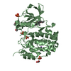
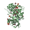
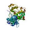
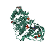
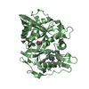
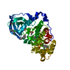
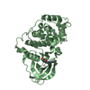
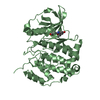
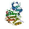
| Title | TLK2 kinase domain from human | ||||||
 Components Components | Serine/threonine-protein kinase tousled-like 2 | ||||||
 Keywords Keywords |  TRANSFERASE / Kinase / ATPgS / chromatin remodelling / DNA replication TRANSFERASE / Kinase / ATPgS / chromatin remodelling / DNA replication | ||||||
| Function / homology |  Function and homology information Function and homology informationregulation of chromatin organization / intermediate filament / negative regulation of proteasomal ubiquitin-dependent protein catabolic process / localization / negative regulation of autophagy / chromosome segregation / cellular response to gamma radiation / chromatin organization / peptidyl-serine phosphorylation / non-specific serine/threonine protein kinase ...regulation of chromatin organization / intermediate filament / negative regulation of proteasomal ubiquitin-dependent protein catabolic process / localization / negative regulation of autophagy / chromosome segregation / cellular response to gamma radiation / chromatin organization / peptidyl-serine phosphorylation / non-specific serine/threonine protein kinase / intracellular signal transduction / protein phosphorylation / protein serine kinase activity / protein serine/threonine kinase activity / DNA damage response / perinuclear region of cytoplasm / nucleoplasm / ATP binding / identical protein binding / nucleusSimilarity search - Function | ||||||
| Biological species |  Homo sapiens (human) Homo sapiens (human) | ||||||
| Method | X-RAY DIFFRACTION / SYNCHROTRON / MOLECULAR REPLACEMENT / Resolution: 2.86 Å | ||||||
 Authors Authors | Mortuza, G.B. / Montoya, G. | ||||||
 Citation Citation | Journal: Nat Commun / Year: 2018 Title: Molecular basis of Tousled-Like Kinase 2 activation. Authors: Mortuza, G.B. / Hermida, D. / Pedersen, A.K. / Segura-Bayona, S. / Lopez-Mendez, B. / Redondo, P. / Ruther, P. / Pozdnyakova, I. / Garrote, A.M. / Munoz, I.G. / Villamor-Paya, M. / Jauset, C. ...Authors: Mortuza, G.B. / Hermida, D. / Pedersen, A.K. / Segura-Bayona, S. / Lopez-Mendez, B. / Redondo, P. / Ruther, P. / Pozdnyakova, I. / Garrote, A.M. / Munoz, I.G. / Villamor-Paya, M. / Jauset, C. / Olsen, J.V. / Stracker, T.H. / Montoya, G. | ||||||
| History |
|
- Structure visualization
| Structure viewer | Molecule: MolmilJmol/JSmol |
|---|
- Downloads & links
-Download
| PDBx/mmCIF format | 5o0y.cif.gz | 134.2 KB | Display | PDBx/mmCIF format |
|---|---|---|---|---|
| PDB format | pdb5o0y.ent.gz | 108.1 KB | Display | PDB format |
| PDBx/mmJSON format | 5o0y.json.gz | Tree view | PDBx/mmJSON format | |
| Others |  Other downloads Other downloads |
-Validation report
| Arichive directory | https://data.pdbj.org/pub/pdb/validation_reports/o0/5o0yftp://data.pdbj.org/pub/pdb/validation_reports/o0/5o0y | HTTPS FTP |
|---|
-Related structure data
| Similar structure data |
|---|

-Links
 PDBj
PDBj
- Assembly
| Deposited unit | 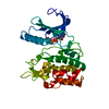
| ||||||||
|---|---|---|---|---|---|---|---|---|---|
| 1 |
| ||||||||
| Unit cell |
|
-Components
| #1: Protein | Mass: 67392.508 Da / Num. of mol.: 1 / Fragment: UNP residues 191-755 Source method: isolated from a genetically manipulated source Source: (gene. exp.) Homo sapiens (human) / Gene: TLK2 / Production host:  Escherichia coli (E. coli) Escherichia coli (E. coli)References: UniProt: Q86UE8, non-specific serine/threonine protein kinase |
|---|---|
| #2: Chemical | ChemComp-AGS /   Mass: 523.247 Da / Num. of mol.: 1 / Source method: obtained synthetically / Formula: C10H16N5O12P3S / Comment: ATP-gamma-S, energy-carrying molecule analogue*YM Mass: 523.247 Da / Num. of mol.: 1 / Source method: obtained synthetically / Formula: C10H16N5O12P3S / Comment: ATP-gamma-S, energy-carrying molecule analogue*YM |
-Experimental details
-Experiment
| Experiment | Method: X-RAY DIFFRACTION / Number of used crystals: 1 |
|---|
- Sample preparation
| Crystal | Density Matthews: 2.47 Å3/Da / Density % sol: 50.3 % |
|---|---|
| Crystal grow | Temperature: 277 K / Method: vapor diffusion, sitting drop Details: 20 mM HEPES pH 7, 2M Li2SO4, 10mM MgCl2 1M Na/K tartrate, 0.1M Tris pH7, 200mM LiSO4 |
-Data collection
| Diffraction | Mean temperature: 100 K |
|---|---|
| Diffraction source | Source: SYNCHROTRON / Site: SLS  / Beamline: X10SA / Wavelength: 0.99987 Å / Beamline: X10SA / Wavelength: 0.99987 Å |
| Detector | Type: DECTRIS PILATUS3 S 6M / Detector: PIXEL / Date: Mar 3, 2014 |
| Radiation | Protocol: SINGLE WAVELENGTH / Monochromatic (M) / Laue (L): M / Scattering type: x-ray |
| Radiation wavelength | Wavelength: 0.99987 Å / Relative weight: 1 |
| Reflection | Resolution: 2.85→72.76 Å / Num. obs: 15761 / % possible obs: 100 % / Redundancy: 2 % / Net I/σ(I): 13.07 |
| Reflection shell | Resolution: 2.857→2.959 Å / Num. unique obs: 15761 / % possible all: 100 |
- Processing
| Software |
| ||||||||||||||||||||||||||||||||||||||||||||||||||||||||||||||||||||||||||||||||||||||||||||||||||||||||||||||||||||||||||||||||||||||||||||||||||||||||||||||||||||||||||||||||||||||
|---|---|---|---|---|---|---|---|---|---|---|---|---|---|---|---|---|---|---|---|---|---|---|---|---|---|---|---|---|---|---|---|---|---|---|---|---|---|---|---|---|---|---|---|---|---|---|---|---|---|---|---|---|---|---|---|---|---|---|---|---|---|---|---|---|---|---|---|---|---|---|---|---|---|---|---|---|---|---|---|---|---|---|---|---|---|---|---|---|---|---|---|---|---|---|---|---|---|---|---|---|---|---|---|---|---|---|---|---|---|---|---|---|---|---|---|---|---|---|---|---|---|---|---|---|---|---|---|---|---|---|---|---|---|---|---|---|---|---|---|---|---|---|---|---|---|---|---|---|---|---|---|---|---|---|---|---|---|---|---|---|---|---|---|---|---|---|---|---|---|---|---|---|---|---|---|---|---|---|---|---|---|---|---|
| Refinement | Method to determine structure: MOLECULAR REPLACEMENT / Resolution: 2.86→89.11 Å / Cor.coef. Fo:Fc: 0.967 / Cor.coef. Fo:Fc free: 0.949 / SU B: 35.731 / SU ML: 0.254 / Cross valid method: THROUGHOUT / ESU R: 0.353 / ESU R Free: 0.244 / Stereochemistry target values: MAXIMUM LIKELIHOOD / Details: HYDROGENS HAVE BEEN ADDED IN THE RIDING POSITIONS
| ||||||||||||||||||||||||||||||||||||||||||||||||||||||||||||||||||||||||||||||||||||||||||||||||||||||||||||||||||||||||||||||||||||||||||||||||||||||||||||||||||||||||||||||||||||||
| Solvent computation | Ion probe radii: 0.9 Å / Shrinkage radii: 0.9 Å / VDW probe radii: 1 Å / Solvent model: MASK | ||||||||||||||||||||||||||||||||||||||||||||||||||||||||||||||||||||||||||||||||||||||||||||||||||||||||||||||||||||||||||||||||||||||||||||||||||||||||||||||||||||||||||||||||||||||
| Displacement parameters | Biso mean: 101.882 Å2
| ||||||||||||||||||||||||||||||||||||||||||||||||||||||||||||||||||||||||||||||||||||||||||||||||||||||||||||||||||||||||||||||||||||||||||||||||||||||||||||||||||||||||||||||||||||||
| Refinement step | Cycle: 1 / Resolution: 2.86→89.11 Å
| ||||||||||||||||||||||||||||||||||||||||||||||||||||||||||||||||||||||||||||||||||||||||||||||||||||||||||||||||||||||||||||||||||||||||||||||||||||||||||||||||||||||||||||||||||||||
| Refine LS restraints |
|