 Movie
Movie Controller

[English] 日本語
 Yorodumi
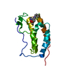
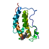
- PDB-5nne: Crystal Structure of the first bromodomain of human BRD4 in compl... -
+ Open data

- Basic information
| Entry | Database: PDB / ID: 5nne | ||||||
|---|---|---|---|---|---|---|---|
| Title | Crystal Structure of the first bromodomain of human BRD4 in complex with a diacetylated TOP2A peptide (K1201ac/K1204ac) | ||||||
 Components Components |
| ||||||
 Keywords Keywords |  TRANSCRIPTION / Bromodomain / complex TRANSCRIPTION / Bromodomain / complex | ||||||
| Function / homology |  Function and homology information Function and homology informationnegative regulation of DNA duplex unwinding / positive regulation of single stranded viral RNA replication via double stranded DNA intermediate / sister chromatid segregation / apoptotic chromosome condensation / DNA topoisomerase type II (double strand cut, ATP-hydrolyzing) complex / resolution of meiotic recombination intermediates / female meiotic nuclear division / DNA ligation / embryonic cleavage / Transcription of E2F targets under negative control by DREAM complex ...negative regulation of DNA duplex unwinding / positive regulation of single stranded viral RNA replication via double stranded DNA intermediate / sister chromatid segregation / apoptotic chromosome condensation / DNA topoisomerase type II (double strand cut, ATP-hydrolyzing) complex / resolution of meiotic recombination intermediates / female meiotic nuclear division / DNA ligation / embryonic cleavage / Transcription of E2F targets under negative control by DREAM complex / DNA topoisomerase type II (double strand cut, ATP-hydrolyzing) activity / DNA binding, bending / DNA topoisomerase (ATP-hydrolysing) / SUMOylation of DNA replication proteins / DNA topological change / RNA polymerase II C-terminal domain binding / chromosome, centromeric region / negative regulation of DNA damage checkpoint / P-TEFb complex binding / negative regulation by host of viral transcription / ATP-dependent activity, acting on DNA / hematopoietic progenitor cell differentiation / condensed chromosome / positive regulation of G2/M transition of mitotic cell cycle / histone reader activity / RNA polymerase II CTD heptapeptide repeat kinase activity / protein kinase C binding / ubiquitin binding / condensed nuclear chromosome / male germ cell nucleus / chromosome segregation / positive regulation of transcription elongation by RNA polymerase II / transcription coregulator activity / lysine-acetylated histone binding / regulation of circadian rhythm / rhythmic process / p53 binding / chromosome / regulation of inflammatory response / positive regulation of canonical NF-kappaB signal transduction / Potential therapeutics for SARS / transcription coactivator activity / transcription cis-regulatory region binding / chromatin remodeling / ribonucleoprotein complex / positive regulation of apoptotic process / protein heterodimerization activity / DNA damage response / chromatin binding / nucleolus / regulation of transcription by RNA polymerase II / positive regulation of DNA-templated transcription / magnesium ion binding / enzyme binding / protein homodimerization activity / positive regulation of transcription by RNA polymerase II / protein-containing complex / DNA binding / RNA binding / nucleoplasm / ATP binding / nucleus / cytoplasmSimilarity search - Function | ||||||
| Biological species |  Homo sapiens (human) Homo sapiens (human) | ||||||
| Method | X-RAY DIFFRACTION / SYNCHROTRON / MOLECULAR REPLACEMENT / Resolution: 1.15 Å | ||||||
 Authors Authors | Filippakopoulos, P. / Picaud, S. / Krojer, T. / von Delft, F. / Arrowsmith, C.H. / Edwards, A.M. / Bountra, C. | ||||||
| Funding support |  United Kingdom, 1items United Kingdom, 1items
| ||||||
 Citation Citation | Journal: Mol. Cell / Year: 2019 Title: Interactome Rewiring Following Pharmacological Targeting of BET Bromodomains. Authors: Lambert, J.P. / Picaud, S. / Fujisawa, T. / Hou, H. / Savitsky, P. / Uuskula-Reimand, L. / Gupta, G.D. / Abdouni, H. / Lin, Z.Y. / Tucholska, M. / Knight, J.D.R. / Gonzalez-Badillo, B. / St- ...Authors: Lambert, J.P. / Picaud, S. / Fujisawa, T. / Hou, H. / Savitsky, P. / Uuskula-Reimand, L. / Gupta, G.D. / Abdouni, H. / Lin, Z.Y. / Tucholska, M. / Knight, J.D.R. / Gonzalez-Badillo, B. / St-Denis, N. / Newman, J.A. / Stucki, M. / Pelletier, L. / Bandeira, N. / Wilson, M.D. / Filippakopoulos, P. / Gingras, A.C. | ||||||
| History |
|
- Structure visualization
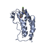
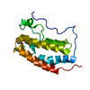
| Structure viewer | Molecule: MolmilJmol/JSmol |
|---|
- Downloads & links
-Download
| PDBx/mmCIF format | 5nne.cif.gz | 78.1 KB | Display | PDBx/mmCIF format |
|---|---|---|---|---|
| PDB format | pdb5nne.ent.gz | 57.5 KB | Display | PDB format |
| PDBx/mmJSON format | 5nne.json.gz | Tree view | PDBx/mmJSON format | |
| Others |  Other downloads Other downloads |
-Validation report
| Arichive directory | https://data.pdbj.org/pub/pdb/validation_reports/nn/5nneftp://data.pdbj.org/pub/pdb/validation_reports/nn/5nne | HTTPS FTP |
|---|
-Related structure data
| Related structure data | 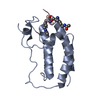 5nncC 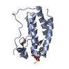 5nndC  5nnfC  5nngC  6g0oC  6g0pC  6g0qC  6g0rC 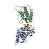 6g0sC 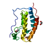 2ossS S: Starting model for refinement C: citing same article ( |
|---|---|
| Similar structure data |


-Links
 PDBj
PDBj


- Assembly
| Deposited unit | 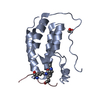
| ||||||||
|---|---|---|---|---|---|---|---|---|---|
| 1 |
| ||||||||
| Unit cell |
|
-Components
| #1: Protein | BRD4 / Protein HUNK1 Mass: 15099.380 Da / Num. of mol.: 1 Source method: isolated from a genetically manipulated source Source: (gene. exp.) Homo sapiens (human) / Gene: BRD4, HUNK1 / Plasmid: pNIC28-Bsa4 / Production host:  Escherichia coli BL21(DE3) (bacteria) / Variant (production host): R3 / References: UniProt: O60885 Escherichia coli BL21(DE3) (bacteria) / Variant (production host): R3 / References: UniProt: O60885 |
|---|---|
| #2: Protein/peptide | Mass: 1325.576 Da / Num. of mol.: 1 / Source method: obtained synthetically Details: TOP2A peptide acetylated at K1201 and K1204. Additional C-terminal TYR added for UV detection Source: (synth.) Homo sapiens (human) / References: UniProt: P11388*PLUS |
| #3: Chemical | ChemComp-EDO / Ethylene glycol  Mass: 62.068 Da / Num. of mol.: 1 / Source method: obtained synthetically / Formula: C2H6O2 Mass: 62.068 Da / Num. of mol.: 1 / Source method: obtained synthetically / Formula: C2H6O2 |
| #4: Water | ChemComp-HOH / Water Mass: 18.015 Da / Num. of mol.: 142 / Source method: isolated from a natural source / Formula: H2O Mass: 18.015 Da / Num. of mol.: 142 / Source method: isolated from a natural source / Formula: H2O |
-Experimental details
-Experiment
| Experiment | Method: X-RAY DIFFRACTION / Number of used crystals: 1 |
|---|
- Sample preparation
| Crystal | Density Matthews: 1.94 Å3/Da / Density % sol: 36.67 % |
|---|---|
| Crystal grow | Temperature: 277 K / Method: vapor diffusion, sitting drop / pH: 7.5 / Details: 20% PEG3350 10% ethylene glycol 0.2M sodium iodide |
-Data collection
| Diffraction | Mean temperature: 100 K |
|---|---|
| Diffraction source | Source: SYNCHROTRON / Site: Diamond / Beamline: I03 / Wavelength: 0.97625 Å |
| Detector | Type: DECTRIS PILATUS 6M / Detector: PIXEL / Date: Dec 15, 2016 |
| Radiation | Protocol: SINGLE WAVELENGTH / Monochromatic (M) / Laue (L): M / Scattering type: x-ray |
| Radiation wavelength | Wavelength: 0.97625 Å / Relative weight: 1 |
| Reflection | Resolution: 1.15→29.1 Å / Num. obs: 46081 / % possible obs: 99 % / Redundancy: 6.9 % / Rmerge(I) obs: 0.037 / Rpim(I) all: 0.015 / Rrim(I) all: 0.015 / Rsym value: 0.037 / Net I/σ(I): 22.5 |
| Reflection shell | Resolution: 1.61→1.7 Å / Redundancy: 4.9 % / Rmerge(I) obs: 0.741 / Mean I/σ(I) obs: 2 / Num. unique all: 2595 / Rsym value: 0.741 / % possible all: 98.2 |
- Processing
| Software |
| ||||||||||||||||||||||||||||||||||||||||||||||||||||||||||||||||||||||||||||||||||||||||||||||||||||||||||||||||||||||||||||||||||||||||||||||||||||||||||||||||||||||||||||||||||||||
|---|---|---|---|---|---|---|---|---|---|---|---|---|---|---|---|---|---|---|---|---|---|---|---|---|---|---|---|---|---|---|---|---|---|---|---|---|---|---|---|---|---|---|---|---|---|---|---|---|---|---|---|---|---|---|---|---|---|---|---|---|---|---|---|---|---|---|---|---|---|---|---|---|---|---|---|---|---|---|---|---|---|---|---|---|---|---|---|---|---|---|---|---|---|---|---|---|---|---|---|---|---|---|---|---|---|---|---|---|---|---|---|---|---|---|---|---|---|---|---|---|---|---|---|---|---|---|---|---|---|---|---|---|---|---|---|---|---|---|---|---|---|---|---|---|---|---|---|---|---|---|---|---|---|---|---|---|---|---|---|---|---|---|---|---|---|---|---|---|---|---|---|---|---|---|---|---|---|---|---|---|---|---|---|
| Refinement | Method to determine structure: MOLECULAR REPLACEMENT Starting model: 2OSS Resolution: 1.15→29.1 Å / Cor.coef. Fo:Fc: 0.975 / Cor.coef. Fo:Fc free: 0.97 / SU B: 0.893 / SU ML: 0.019 / Cross valid method: THROUGHOUT / ESU R: 0.032 / ESU R Free: 0.033 / Details: HYDROGENS HAVE BEEN ADDED IN THE RIDING POSITIONS
| ||||||||||||||||||||||||||||||||||||||||||||||||||||||||||||||||||||||||||||||||||||||||||||||||||||||||||||||||||||||||||||||||||||||||||||||||||||||||||||||||||||||||||||||||||||||
| Solvent computation | Ion probe radii: 0.8 Å / Shrinkage radii: 0.8 Å / VDW probe radii: 1.2 Å | ||||||||||||||||||||||||||||||||||||||||||||||||||||||||||||||||||||||||||||||||||||||||||||||||||||||||||||||||||||||||||||||||||||||||||||||||||||||||||||||||||||||||||||||||||||||
| Displacement parameters | Biso mean: 18.087 Å2
| ||||||||||||||||||||||||||||||||||||||||||||||||||||||||||||||||||||||||||||||||||||||||||||||||||||||||||||||||||||||||||||||||||||||||||||||||||||||||||||||||||||||||||||||||||||||
| Refinement step | Cycle: 1 / Resolution: 1.15→29.1 Å
| ||||||||||||||||||||||||||||||||||||||||||||||||||||||||||||||||||||||||||||||||||||||||||||||||||||||||||||||||||||||||||||||||||||||||||||||||||||||||||||||||||||||||||||||||||||||
| Refine LS restraints |
|