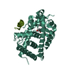
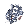
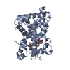
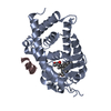
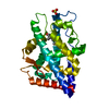
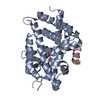
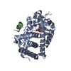
[English] 日本語
Yorodumi
- PDB-5nky: Structure-activity relationship study of vitamin D analogs with o... -

+
Open data


ID or keywords:

Loading...

-
Basic information

Entry
Database: PDB / ID: 5nky
TitleStructure-activity relationship study of vitamin D analogs with oxolane group in their side chain
Components
  • SRC1
  • Vitamin D3 receptor A
KeywordsGENE REGULATION
Function / homology
Function and homology information


heart jogging / Vitamin D (calciferol) metabolism / : / SUMOylation of intracellular receptors / vitamin D binding / calcitriol binding / lithocholic acid binding / labyrinthine layer morphogenesis / regulation of thyroid hormone mediated signaling pathway / positive regulation of transcription from RNA polymerase II promoter by galactose ...heart jogging / Vitamin D (calciferol) metabolism / : / SUMOylation of intracellular receptors / vitamin D binding / calcitriol binding / lithocholic acid binding / labyrinthine layer morphogenesis / regulation of thyroid hormone mediated signaling pathway / positive regulation of transcription from RNA polymerase II promoter by galactose / positive regulation of female receptivity / hematopoietic stem cell proliferation / hypothalamus development / male mating behavior / NR1H2 & NR1H3 regulate gene expression to control bile acid homeostasis / heart looping / estrous cycle / cellular response to Thyroglobulin triiodothyronine / Synthesis of bile acids and bile salts / calcium ion homeostasis / Endogenous sterols / Synthesis of bile acids and bile salts via 27-hydroxycholesterol / Synthesis of bile acids and bile salts via 7alpha-hydroxycholesterol / nuclear retinoid X receptor binding / NR1H3 & NR1H2 regulate gene expression linked to cholesterol transport and efflux / response to retinoic acid / histone acetyltransferase activity / Recycling of bile acids and salts / regulation of cellular response to insulin stimulus / histone acetyltransferase / cellular response to hormone stimulus / positive regulation of adipose tissue development / peroxisome proliferator activated receptor signaling pathway / RORA activates gene expression / positive regulation of neuron differentiation / lactation / Regulation of lipid metabolism by PPARalpha / cerebellum development / ossification / BMAL1:CLOCK,NPAS2 activates circadian gene expression / nuclear receptor coactivator activity / SUMOylation of transcription cofactors / Activation of gene expression by SREBF (SREBP) / response to progesterone / nuclear estrogen receptor binding / nuclear receptor binding / hippocampus development / RNA polymerase II transcription regulatory region sequence-specific DNA binding / Heme signaling / mRNA transcription by RNA polymerase II / Transcriptional activation of mitochondrial biogenesis / PPARA activates gene expression / Cytoprotection by HMOX1 / cerebral cortex development / Transcriptional regulation of white adipocyte differentiation / RNA polymerase II transcription regulator complex / male gonad development / nuclear receptor activity / Circadian Clock / response to estradiol / HATs acetylate histones / Estrogen-dependent gene expression / transcription regulator complex / cell differentiation / transcription coactivator activity / protein dimerization activity / positive regulation of apoptotic process / RNA polymerase II cis-regulatory region sequence-specific DNA binding / DNA-templated transcription / chromatin binding / chromatin / regulation of DNA-templated transcription / positive regulation of DNA-templated transcription / negative regulation of transcription by RNA polymerase II / positive regulation of transcription by RNA polymerase II / protein-containing complex / zinc ion binding / nucleoplasm / nucleus / plasma membrane / cytosol / cytoplasm
Similarity search - Function
Vitamin D receptor / VDR, DNA-binding domain / Nuclear receptor coactivator 1 / Nuclear receptor coactivator, DUF1518 / Nuclear receptor coactivator, Ncoa-type, interlocking / Nuclear receptor coactivator, Ncoa-type, interlocking domain superfamily / Nuclear receptor coactivator, DUF1518 / Nuclear receptor coactivator / DUF1518 / Nuclear receptor coactivator, receptor-binding domain ...Vitamin D receptor / VDR, DNA-binding domain / Nuclear receptor coactivator 1 / Nuclear receptor coactivator, DUF1518 / Nuclear receptor coactivator, Ncoa-type, interlocking / Nuclear receptor coactivator, Ncoa-type, interlocking domain superfamily / Nuclear receptor coactivator, DUF1518 / Nuclear receptor coactivator / DUF1518 / Nuclear receptor coactivator, receptor-binding domain / Nuclear receptor coactivator / Steroid receptor coactivator / Unstructured region on nuclear receptor coactivator protein / PAS domain / Nuclear receptor coactivator, interlocking / Helix-loop-helix DNA-binding domain superfamily / helix loop helix domain / Myc-type, basic helix-loop-helix (bHLH) domain / Myc-type, basic helix-loop-helix (bHLH) domain profile. / PAS fold / PAS fold / PAS domain / PAS repeat profile. / PAS domain / PAS domain superfamily / Retinoid X Receptor / Retinoid X Receptor / Nuclear hormone receptor / Nuclear hormones receptors DNA-binding region signature. / Zinc finger, nuclear hormone receptor-type / Zinc finger, C4 type (two domains) / Nuclear hormone receptors DNA-binding domain profile. / c4 zinc finger in nuclear hormone receptors / Nuclear hormone receptor, ligand-binding domain / Nuclear hormone receptor-like domain superfamily / Ligand-binding domain of nuclear hormone receptor / Nuclear receptor (NR) ligand-binding (LBD) domain profile. / Ligand binding domain of hormone receptors / Zinc finger, NHR/GATA-type / Orthogonal Bundle / Mainly Alpha
Similarity search - Domain/homology
Chem-91W / Nuclear receptor coactivator 1 / Vitamin D3 receptor A
Similarity search - Component
Biological speciesDanio rerio (zebrafish)
Homo sapiens (human)
MethodX-RAY DIFFRACTION / SYNCHROTRON / MOLECULAR REPLACEMENT / Resolution: 2.096 Å
AuthorsRochel, N. / Belorusova, A.Y.
Funding support France, 2items
OrganizationGrant numberCountry
French National Research AgencyANR-13-BSV8-0024-01 France
FRM(FRM: FDT20140930978 France
CitationJournal: Eur J Med Chem / Year: 2017
Title: Structure-activity relationship study of vitamin D analogs with oxolane group in their side chain.
Authors: Belorusova, A.Y. / Martinez, A. / Gandara, Z. / Gomez, G. / Fall, Y. / Rochel, N.
History
DepositionApr 3, 2017Deposition site: PDBE / Processing site: PDBE
Revision 1.0May 24, 2017Provider: repository / Type: Initial release
Revision 1.1Sep 6, 2017Group: Author supporting evidence / Category: pdbx_audit_support / Item: _pdbx_audit_support.funding_organization
Revision 1.2Feb 24, 2021Group: Derived calculations
Category: pdbx_struct_assembly / pdbx_struct_assembly_gen ...pdbx_struct_assembly / pdbx_struct_assembly_gen / pdbx_struct_assembly_prop / pdbx_struct_oper_list
Revision 1.3Jan 17, 2024Group: Advisory / Data collection ...Advisory / Data collection / Database references / Refinement description
Category: chem_comp_atom / chem_comp_bond ...chem_comp_atom / chem_comp_bond / database_2 / pdbx_initial_refinement_model / pdbx_unobs_or_zero_occ_atoms
Item: _database_2.pdbx_DOI / _database_2.pdbx_database_accession

-
Structure visualization

Structure viewerMolecule:
MolmilJmol/JSmol

Downloads & links

-
Assembly

Deposited unit
A: Vitamin D3 receptor A
B: SRC1
hetero molecules


Theoretical massNumber of molelcules
Total (without water)36,2813
Polymers35,8372
Non-polymers4451
Water27015
1
A: Vitamin D3 receptor A
B: SRC1
hetero molecules

A: Vitamin D3 receptor A
B: SRC1
hetero molecules


Theoretical massNumber of molelcules
Total (without water)72,5636
Polymers71,6734
Non-polymers8892
Water362
TypeNameSymmetry operationNumber
identity operation1_555x,y,z1
crystal symmetry operation12_564x,x-y+1,-z-1/61
Buried area3980 Å2
ΔGint-30 kcal/mol
Surface area21750 Å2
MethodPISA
Unit cell
Length a, b, c (Å)66.191, 66.191, 261.698
Angle α, β, γ (deg.)90.00, 90.00, 120.00
Int Tables number179
Space group name H-MP6522

-
Components

#1: Protein Vitamin D3 receptor A / VDR-A / 1 / 25-dihydroxyvitamin D3 receptor A / Nuclear receptor subfamily 1 group I member 1-A


Mass: 34060.672 Da / Num. of mol.: 1
Source method: isolated from a genetically manipulated source
Source: (gene. exp.) Danio rerio (zebrafish) / Gene: vdra, nr1i1a, vdr / Production host: Escherichia coli (E. coli) / References: UniProt: Q9PTN2
#2: Protein/peptide SRC1 /


Mass: 1776.072 Da / Num. of mol.: 1 / Source method: obtained synthetically / Source: (synth.) Homo sapiens (human) / References: UniProt: Q15788*PLUS
#3: Chemical ChemComp-91W / (1~{R},3~{S},5~{Z})-5-[(2~{E})-2-[(1~{R},3~{a}~{S},7~{a}~{R})-7~{a}-methyl-1-[(1~{S})-1-[(2~{R},5~{S})-5-(2-oxidanylpropan-2-yl)oxolan-2-yl]ethyl]-2,3,3~{a},5,6,7-hexahydro-1~{H}-inden-4-ylidene]ethylidene]-4-methylidene-cyclohexane-1,3-diol


Mass: 444.647 Da / Num. of mol.: 1 / Source method: obtained synthetically / Formula: C28H44O4
#4: Water ChemComp-HOH / water / Water


Mass: 18.015 Da / Num. of mol.: 15 / Source method: isolated from a natural source / Formula: H2O

-
Experimental details

-
Experiment

ExperimentMethod: X-RAY DIFFRACTION / Number of used crystals: 1

-
Sample preparation

CrystalDensity Matthews: 2.31 Å3/Da / Density % sol: 46.73 %
Crystal growTemperature: 290 K / Method: vapor diffusion, sitting drop / pH: 7 / Details: 0.1 M BTP pH 7.0, 3 M NaOAc

-
Data collection

DiffractionMean temperature: 100 K
Diffraction sourceSource: SYNCHROTRON / Site: ESRF / Beamline: ID29 / Wavelength: 0.9786 Å
DetectorType: DECTRIS PILATUS3 S 6M / Detector: PIXEL / Date: May 28, 2014
RadiationProtocol: SINGLE WAVELENGTH / Monochromatic (M) / Laue (L): M / Scattering type: x-ray
Radiation wavelengthWavelength: 0.9786 Å / Relative weight: 1
ReflectionResolution: 2.096→50 Å / Num. obs: 17559 / % possible obs: 84 % / Redundancy: 15.3 % / Rsym value: 0.087 / Net I/σ(I): 27
Reflection shellResolution: 2.096→2.14 Å / Redundancy: 8.8 % / Rsym value: 0.553 / % possible all: 31

-
Processing

Software
NameVersionClassification
PHENIXdev_1980refinement
HKL-2000data reduction
HKL-2000data scaling
PHENIXphasing
RefinementMethod to determine structure: MOLECULAR REPLACEMENT
Starting model: 2HC4
Resolution: 2.096→43.616 Å / SU ML: 0.26 / Cross valid method: FREE R-VALUE / σ(F): 1.34 / Phase error: 35.45
RfactorNum. reflection% reflectionSelection details
Rfree0.2463 859 4.91 %RANDOM SELECTION
Rwork0.2262 ---
obs0.2272 17505 83.36 %-
Solvent computationShrinkage radii: 0.9 Å / VDW probe radii: 1.11 Å
Refinement stepCycle: LAST / Resolution: 2.096→43.616 Å
ProteinNucleic acidLigandSolventTotal
Num. atoms1985 0 32 15 2032
Refine LS restraints
Refine-IDTypeDev idealNumber
X-RAY DIFFRACTIONf_bond_d0.0072058
X-RAY DIFFRACTIONf_angle_d0.9932780
X-RAY DIFFRACTIONf_dihedral_angle_d14.738799
X-RAY DIFFRACTIONf_chiral_restr0.041318
X-RAY DIFFRACTIONf_plane_restr0.005353
LS refinement shell
Resolution (Å)Rfactor RfreeNum. reflection RfreeRfactor RworkNum. reflection RworkRefine-ID% reflection obs (%)
2.0961-2.22740.4189550.28581126X-RAY DIFFRACTION35
2.2274-2.39940.313930.30652164X-RAY DIFFRACTION66
2.3994-2.64080.3091620.28533170X-RAY DIFFRACTION97
2.6408-3.02280.26721620.28523295X-RAY DIFFRACTION99
3.0228-3.80810.281920.2433308X-RAY DIFFRACTION100
3.8081-43.62580.2091950.1913583X-RAY DIFFRACTION100

+
About Yorodumi

-
News

-
Feb 9, 2022. New format data for meta-information of EMDB entries

New format data for meta-information of EMDB entries

  • Version 3 of the EMDB header file is now the official format.
  • The previous official version 1.9 will be removed from the archive.

Related info.:EMDB header

External links:wwPDB to switch to version 3 of the EMDB data model

-
Aug 12, 2020. Covid-19 info

Covid-19 info

URL: https://pdbj.org/emnavi/covid19.php

New page: Covid-19 featured information page in EM Navigator.

Related info.:Covid-19 info / Mar 5, 2020. Novel coronavirus structure data

+
Mar 5, 2020. Novel coronavirus structure data

Novel coronavirus structure data

Related info.:Yorodumi Speices / Aug 12, 2020. Covid-19 info

External links:COVID-19 featured content - PDBj / Molecule of the Month (242):Coronavirus Proteases

+
Jan 31, 2019. EMDB accession codes are about to change! (news from PDBe EMDB page)

EMDB accession codes are about to change! (news from PDBe EMDB page)

  • The allocation of 4 digits for EMDB accession codes will soon come to an end. Whilst these codes will remain in use, new EMDB accession codes will include an additional digit and will expand incrementally as the available range of codes is exhausted. The current 4-digit format prefixed with “EMD-” (i.e. EMD-XXXX) will advance to a 5-digit format (i.e. EMD-XXXXX), and so on. It is currently estimated that the 4-digit codes will be depleted around Spring 2019, at which point the 5-digit format will come into force.
  • The EM Navigator/Yorodumi systems omit the EMD- prefix.

Related info.:Q: What is EMD? / ID/Accession-code notation in Yorodumi/EM Navigator

External links:EMDB Accession Codes are Changing Soon! / Contact to PDBj

+
Jul 12, 2017. Major update of PDB

Major update of PDB

  • wwPDB released updated PDB data conforming to the new PDBx/mmCIF dictionary.
  • This is a major update changing the version number from 4 to 5, and with Remediation, in which all the entries are updated.
  • In this update, many items about electron microscopy experimental information are reorganized (e.g. em_software).
  • Now, EM Navigator and Yorodumi are based on the updated data.

External links:wwPDB Remediation / Enriched Model Files Conforming to OneDep Data Standards Now Available in the PDB FTP Archive

-
Yorodumi

Thousand views of thousand structures

  • Yorodumi is a browser for structure data from EMDB, PDB, SASBDB, etc.
  • This page is also the successor to EM Navigator detail page, and also detail information page/front-end page for Omokage search.
  • The word "yorodu" (or yorozu) is an old Japanese word meaning "ten thousand". "mi" (miru) is to see.

Related info.:EMDB / PDB / SASBDB / Comparison of 3 databanks / Yorodumi Search / Aug 31, 2016. New EM Navigator & Yorodumi / Yorodumi Papers / Jmol/JSmol / Function and homology information / Changes in new EM Navigator and Yorodumi

Read more