 Movie
Movie Controller

[English] 日本語
 Yorodumi
- PDB-5mrj: Crystal structure of Endo-1,4-beta-xylanase-like protein from Acr... -
+ Open data

- Basic information
| Entry | Database: PDB / ID: 5mrj | ||||||
|---|---|---|---|---|---|---|---|
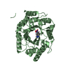
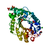
| Title | Crystal structure of Endo-1,4-beta-xylanase-like protein from Acremonium chrysogenum | ||||||
 Components Components | Beta-xylanase Xylanase Xylanase | ||||||
 Keywords Keywords | HYDROLASE / xylanase-like protein / Endo-1 / 4-beta-xylanase-like protein | ||||||
| Function / homology |  Function and homology informationcellulose binding / endo-1,4-beta-xylanase activity / endo-1,4-beta-xylanase / xylan catabolic process / extracellular region Function and homology informationcellulose binding / endo-1,4-beta-xylanase activity / endo-1,4-beta-xylanase / xylan catabolic process / extracellular regionSimilarity search - Function | ||||||
| Biological species |  Acremonium chrysogenum (fungus) Acremonium chrysogenum (fungus) | ||||||
| Method | X-RAY DIFFRACTION / SYNCHROTRON / MOLECULAR REPLACEMENT / Resolution: 2.7 Å | ||||||
 Authors Authors | Gabdulkhakov, A. / Tishchenko, S. / Lisov, A. / Leontievsky, A. | ||||||
 Citation Citation | Journal: To Be Published Title: Crystal structure of Endo-1,4-beta-xylanase-like protein from Acremonium chrysogenum Authors: Gabdulkhakov, A. / Tishchenko, S. / Lisov, A. / Leontievsky, A. | ||||||
| History |
|
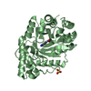
- Structure visualization
| Structure viewer | Molecule: MolmilJmol/JSmol |
|---|
- Downloads & links
-Download
| PDBx/mmCIF format | 5mrj.cif.gz | 131.5 KB | Display | PDBx/mmCIF format |
|---|---|---|---|---|
| PDB format | pdb5mrj.ent.gz | 101.4 KB | Display | PDB format |
| PDBx/mmJSON format | 5mrj.json.gz | Tree view | PDBx/mmJSON format | |
| Others |  Other downloads Other downloads |
-Validation report
| Arichive directory | https://data.pdbj.org/pub/pdb/validation_reports/mr/5mrjftp://data.pdbj.org/pub/pdb/validation_reports/mr/5mrj | HTTPS FTP |
|---|
-Related structure data
| Related structure data |  1b30S S: Starting model for refinement |
|---|---|
| Similar structure data |


-Links
 PDBj
PDBj
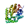
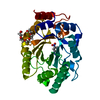
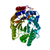
- Assembly
| Deposited unit | 
| ||||||||
|---|---|---|---|---|---|---|---|---|---|
| 1 | 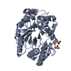
| ||||||||
| 2 | 
| ||||||||
| Unit cell |
|
-Components
| #1: Protein | Xylanase / Endo-1 / 4-beta-xylanase-like protein Mass: 43170.207 Da / Num. of mol.: 2 Source method: isolated from a genetically manipulated source Source: (gene. exp.) Acremonium chrysogenum (strain ATCC 11550 / CBS 779.69 / DSM 880 / JCM 23072 / IMI 49137) (fungus)Strain: ATCC 11550 / CBS 779.69 / DSM 880 / JCM 23072 / IMI 49137 Gene: ACRE_072080 / Plasmid: pPic9m / Production host: Komagataella phaffii GS115 (fungus) / References: UniProt: A0A086SY89, endo-1,4-beta-xylanase#2: Chemical | Sulfate  Mass: 96.063 Da / Num. of mol.: 3 / Source method: obtained synthetically / Formula: SO4 Mass: 96.063 Da / Num. of mol.: 3 / Source method: obtained synthetically / Formula: SO4#3: Water | ChemComp-HOH / | Water Mass: 18.015 Da / Num. of mol.: 41 / Source method: isolated from a natural source / Formula: H2O Mass: 18.015 Da / Num. of mol.: 41 / Source method: isolated from a natural source / Formula: H2O |
|---|
-Experimental details
-Experiment
| Experiment | Method: X-RAY DIFFRACTION / Number of used crystals: 1 |
|---|
- Sample preparation
| Crystal | Density Matthews: 3.37 Å3/Da / Density % sol: 63.55 % |
|---|---|
| Crystal grow | Temperature: 295 K / Method: vapor diffusion, hanging drop / pH: 5.5 Details: 15% PEG 4K, 50 mM Sodium Citrate, 20 mM Sodium chloride, 20 mM Ammonium Sulfate |
-Data collection
| Diffraction | Mean temperature: 100 K |
|---|---|
| Diffraction source | Source: SYNCHROTRON / Site: BESSY  / Beamline: 14.1 / Wavelength: 0.9677 Å / Beamline: 14.1 / Wavelength: 0.9677 Å |
| Detector | Type: DECTRIS PILATUS3 S 6M / Detector: PIXEL / Date: Nov 19, 2016 |
| Radiation | Protocol: SINGLE WAVELENGTH / Monochromatic (M) / Laue (L): M / Scattering type: x-ray |
| Radiation wavelength | Wavelength: 0.9677 Å / Relative weight: 1 |
| Reflection | Resolution: 2.64→50 Å / Num. obs: 25746 / % possible obs: 94.3 % / Redundancy: 3.95 % / Net I/σ(I): 21 |
| Reflection shell | Resolution: 2.64→2.8 Å / Redundancy: 3.71 % / % possible all: 96.6 |
- Processing
| Software |
| ||||||||||||||||||||||||||||||||||||||||||||||||||||||||||||||||||||||||||||||||||||||||||||||||||||||||||||||||||||||||||||||
|---|---|---|---|---|---|---|---|---|---|---|---|---|---|---|---|---|---|---|---|---|---|---|---|---|---|---|---|---|---|---|---|---|---|---|---|---|---|---|---|---|---|---|---|---|---|---|---|---|---|---|---|---|---|---|---|---|---|---|---|---|---|---|---|---|---|---|---|---|---|---|---|---|---|---|---|---|---|---|---|---|---|---|---|---|---|---|---|---|---|---|---|---|---|---|---|---|---|---|---|---|---|---|---|---|---|---|---|---|---|---|---|---|---|---|---|---|---|---|---|---|---|---|---|---|---|---|---|
| Refinement | Method to determine structure: MOLECULAR REPLACEMENT Starting model: 1b30 Resolution: 2.7→46.809 Å / SU ML: 0.37 / Cross valid method: FREE R-VALUE / σ(F): 0.09 / Phase error: 32.56
| ||||||||||||||||||||||||||||||||||||||||||||||||||||||||||||||||||||||||||||||||||||||||||||||||||||||||||||||||||||||||||||||
| Solvent computation | Shrinkage radii: 0.9 Å / VDW probe radii: 1.11 Å | ||||||||||||||||||||||||||||||||||||||||||||||||||||||||||||||||||||||||||||||||||||||||||||||||||||||||||||||||||||||||||||||
| Refinement step | Cycle: LAST / Resolution: 2.7→46.809 Å
| ||||||||||||||||||||||||||||||||||||||||||||||||||||||||||||||||||||||||||||||||||||||||||||||||||||||||||||||||||||||||||||||
| Refine LS restraints |
| ||||||||||||||||||||||||||||||||||||||||||||||||||||||||||||||||||||||||||||||||||||||||||||||||||||||||||||||||||||||||||||||
| LS refinement shell |
|
