 Movie
Movie Controller


+ Open data

- Basic information
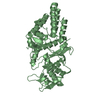
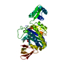
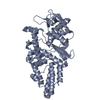
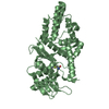
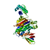
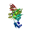
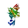
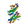
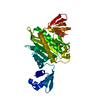
| Entry | Database: PDB / ID: 5mnj | |||||||||
|---|---|---|---|---|---|---|---|---|---|---|
| Title | Structure of MDM2-MDMX-UbcH5B-ubiquitin complex | |||||||||
 Components Components |
| |||||||||
 Keywords Keywords |  LIGASE / Ubiquitin ligase LIGASE / Ubiquitin ligase | |||||||||
| Function / homology |  Function and homology information Function and homology informationcellular response to vitamin B1 / response to formaldehyde / response to water-immersion restraint stress / (E3-independent) E2 ubiquitin-conjugating enzyme / response to ether / traversing start control point of mitotic cell cycle / negative regulation of intrinsic apoptotic signaling pathway by p53 class mediator / negative regulation of signal transduction by p53 class mediator / fibroblast activation / atrial septum development ...cellular response to vitamin B1 / response to formaldehyde / response to water-immersion restraint stress / (E3-independent) E2 ubiquitin-conjugating enzyme / response to ether / traversing start control point of mitotic cell cycle / negative regulation of intrinsic apoptotic signaling pathway by p53 class mediator / negative regulation of signal transduction by p53 class mediator / fibroblast activation / atrial septum development / receptor serine/threonine kinase binding / Trafficking of AMPA receptors / heart valve development / positive regulation of vascular associated smooth muscle cell migration / hypothalamus gonadotrophin-releasing hormone neuron development / peroxisome proliferator activated receptor binding / response to iron ion / negative regulation of protein processing / response to steroid hormone / SUMO transferase activity / NEDD8 ligase activity / female meiosis I / positive regulation of protein monoubiquitination / mitochondrion transport along microtubule / AKT phosphorylates targets in the cytosol / cellular response to peptide hormone stimulus / atrioventricular valve morphogenesis / E2 ubiquitin-conjugating enzyme / fat pad development / ventricular septum development / endocardial cushion morphogenesis / female gonad development / cellular response to alkaloid / positive regulation of muscle cell differentiation / SUMOylation of ubiquitinylation proteins / seminiferous tubule development / blood vessel development / regulation of protein catabolic process / male meiosis I / cardiac septum morphogenesis / ubiquitin conjugating enzyme activity / Constitutive Signaling by AKT1 E17K in Cancer / negative regulation of DNA damage response, signal transduction by p53 class mediator / response to magnesium ion / protein sumoylation / ligase activity / SUMOylation of transcription factors / positive regulation of intrinsic apoptotic signaling pathway by p53 class mediator / protein localization to nucleus / cellular response to actinomycin D / DNA damage response, signal transduction by p53 class mediator / cellular response to UV-C / protein K48-linked ubiquitination / blood vessel remodeling / cellular response to estrogen stimulus / protein autoubiquitination / regulation of proteasomal protein catabolic process / ribonucleoprotein complex binding / ubiquitin ligase complex / energy homeostasis / regulation of neuron apoptotic process / DNA damage response, signal transduction by p53 class mediator resulting in cell cycle arrest / Maturation of protein E / Maturation of protein E / ER Quality Control Compartment (ERQC) / Myoclonic epilepsy of Lafora / FLT3 signaling by CBL mutants / Prevention of phagosomal-lysosomal fusion / IRAK2 mediated activation of TAK1 complex / Alpha-protein kinase 1 signaling pathway / Glycogen synthesis / IRAK1 recruits IKK complex / IRAK1 recruits IKK complex upon TLR7/8 or 9 stimulation / Membrane binding and targetting of GAG proteins / Constitutive Signaling by NOTCH1 HD Domain Mutants / NOTCH2 Activation and Transmission of Signal to the Nucleus / positive regulation of vascular associated smooth muscle cell proliferation / Endosomal Sorting Complex Required For Transport (ESCRT) / IRAK2 mediated activation of TAK1 complex upon TLR7/8 or 9 stimulation / Regulation of FZD by ubiquitination / PTK6 Regulates RTKs and Their Effectors AKT1 and DOK1 / Negative regulation of FLT3 / TICAM1,TRAF6-dependent induction of TAK1 complex / TICAM1-dependent activation of IRF3/IRF7 / APC/C:Cdc20 mediated degradation of Cyclin B / Downregulation of ERBB4 signaling / p75NTR recruits signalling complexes / TRAF6 mediated IRF7 activation in TLR7/8 or 9 signaling / APC-Cdc20 mediated degradation of Nek2A / PINK1-PRKN Mediated Mitophagy / transcription repressor complex / TRAF6-mediated induction of TAK1 complex within TLR4 complex / InlA-mediated entry of Listeria monocytogenes into host cells / Pexophagy / Regulation of innate immune responses to cytosolic DNA / NPAS4 regulates expression of target genes / VLDLR internalisation and degradation / regulation of heart rate / Downregulation of ERBB2:ERBB3 signaling / NRIF signals cell death from the nucleusSimilarity search - Function | |||||||||
| Biological species |  Homo sapiens (human) Homo sapiens (human) | |||||||||
| Method | X-RAY DIFFRACTION / SYNCHROTRON / MOLECULAR REPLACEMENT / Resolution: 2.16 Å | |||||||||
 Authors Authors | Klejnot, M. / Huang, D.T. | |||||||||
| Funding support |  United Kingdom, 2items United Kingdom, 2items
| |||||||||
 Citation Citation | Journal: Nat. Struct. Mol. Biol. / Year: 2017 Title: Structural analysis of MDM2 RING separates degradation from regulation of p53 transcription activity. Authors: Nomura, K. / Klejnot, M. / Kowalczyk, D. / Hock, A.K. / Sibbet, G.J. / Vousden, K.H. / Huang, D.T. | |||||||||
| History |
|
- Structure visualization
| Structure viewer | Molecule: MolmilJmol/JSmol |
|---|
- Downloads & links
-Download
| PDBx/mmCIF format | 5mnj.cif.gz | 150.8 KB | Display | PDBx/mmCIF format |
|---|---|---|---|---|
| PDB format | pdb5mnj.ent.gz | 115.2 KB | Display | PDB format |
| PDBx/mmJSON format | 5mnj.json.gz | Tree view | PDBx/mmJSON format | |
| Others |  Other downloads Other downloads |
-Validation report
| Arichive directory | https://data.pdbj.org/pub/pdb/validation_reports/mn/5mnjftp://data.pdbj.org/pub/pdb/validation_reports/mn/5mnj | HTTPS FTP |
|---|
-Related structure data


-Links
 PDBj
PDBj


- Assembly
| Deposited unit | 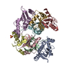
| ||||||||
|---|---|---|---|---|---|---|---|---|---|
| 1 | 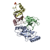
| ||||||||
| 2 | 
| ||||||||
| Unit cell |
|
-Components
-Protein , 4 types, 8 molecules AEBFCGDH
| #1: Protein | Mass: 16851.381 Da / Num. of mol.: 2 / Mutation: S22R, C85K Source method: isolated from a genetically manipulated source Details: K85 in Chains A and E form isopeptide linkage with the carbonyl carbon of G76 in Chains B and F, respectively. Source: (gene. exp.) Homo sapiens (human) / Gene: UBE2D2, PUBC1, UBC4, UBC5B, UBCH4, UBCH5B / Production host:  Escherichia coli (E. coli) Escherichia coli (E. coli)References: UniProt: P62837, E2 ubiquitin-conjugating enzyme, (E3-independent) E2 ubiquitin-conjugating enzyme#2: Protein | Mass: 8922.141 Da / Num. of mol.: 2 Source method: isolated from a genetically manipulated source Details: gsggs linker at the N-terminus resulted from cloning. G76 in chain B is covalently linked to K85 side chain in Chain A. Source: (gene. exp.) Homo sapiens (human) / Gene: UBB / Production host: Escherichia coli (E. coli) / References: UniProt: P0CG47#3: Protein | Mass: 9668.399 Da / Num. of mol.: 2 Source method: isolated from a genetically manipulated source Details: Contains N-terminal His-tag followed by TEV protease cleavage site that was not removed during purification. MDM2 contains 428-491. Source: (gene. exp.) Homo sapiens (human) / Gene: MDM2 / Production host: Escherichia coli (E. coli)References: UniProt: Q00987, Ligases; Forming carbon-nitrogen bonds; Acid-amino-acid ligases (peptide synthases)#4: Protein | Mass: 7207.716 Da / Num. of mol.: 2 Source method: isolated from a genetically manipulated source Details: MDMX contains 427-490 / Source: (gene. exp.) Homo sapiens (human) / Gene: MDM4, MDMX / Production host: Escherichia coli (E. coli) / References: UniProt: O15151 |
|---|
-Non-polymers , 3 types, 87 molecules 


| #5: Chemical | ChemComp-ZN /  Mass: 65.409 Da / Num. of mol.: 8 / Source method: obtained synthetically / Formula: Zn Mass: 65.409 Da / Num. of mol.: 8 / Source method: obtained synthetically / Formula: Zn#6: Chemical | Sulfate Mass: 96.063 Da / Num. of mol.: 2 / Source method: obtained synthetically / Formula: SO4 Mass: 96.063 Da / Num. of mol.: 2 / Source method: obtained synthetically / Formula: SO4#7: Water | ChemComp-HOH / | WaterMass: 18.015 Da / Num. of mol.: 77 / Source method: isolated from a natural source / Formula: H2O |
|---|
-Experimental details
-Experiment
| Experiment | Method: X-RAY DIFFRACTION / Number of used crystals: 1 |
|---|
- Sample preparation
| Crystal | Density Matthews: 2.31 Å3/Da / Density % sol: 46.84 % |
|---|---|
| Crystal grow | Temperature: 292 K / Method: vapor diffusion, hanging drop Details: 0.1 M Tris-HCl, pH 8.5, 0.175 M Li2SO4 and 16-20 %(v/v) PEG 3350 |
-Data collection
| Diffraction | Mean temperature: 100 K |
|---|---|
| Diffraction source | Source: SYNCHROTRON / Site: Diamond / Beamline: I24 / Wavelength: 0.97879 Å |
| Detector | Type: DECTRIS PILATUS 6M / Detector: PIXEL / Date: Feb 21, 2013 |
| Radiation | Protocol: SINGLE WAVELENGTH / Monochromatic (M) / Laue (L): M / Scattering type: x-ray |
| Radiation wavelength | Wavelength: 0.97879 Å / Relative weight: 1 |
| Reflection | Resolution: 2.16→50.5 Å / Num. obs: 37881 / % possible obs: 95.3 % / Redundancy: 3.2 % / Net I/σ(I): 6.8 |
- Processing
| Software |
| |||||||||||||||||||||||||||||||||||||||||||||||||||||||||||||||||||||||||||||||||||||||||||||||||||||||||
|---|---|---|---|---|---|---|---|---|---|---|---|---|---|---|---|---|---|---|---|---|---|---|---|---|---|---|---|---|---|---|---|---|---|---|---|---|---|---|---|---|---|---|---|---|---|---|---|---|---|---|---|---|---|---|---|---|---|---|---|---|---|---|---|---|---|---|---|---|---|---|---|---|---|---|---|---|---|---|---|---|---|---|---|---|---|---|---|---|---|---|---|---|---|---|---|---|---|---|---|---|---|---|---|---|---|---|
| Refinement | Method to determine structure: MOLECULAR REPLACEMENT Starting model: 3ZNI and 3VJF Resolution: 2.16→50.49 Å / SU ML: 0.59 / Cross valid method: FREE R-VALUE / σ(F): 1.96 / Phase error: 30.25
| |||||||||||||||||||||||||||||||||||||||||||||||||||||||||||||||||||||||||||||||||||||||||||||||||||||||||
| Solvent computation | Shrinkage radii: 0.95 Å / VDW probe radii: 1.2 Å / Bsol: 46.114 Å2 / ksol: 0.336 e/Å3 | |||||||||||||||||||||||||||||||||||||||||||||||||||||||||||||||||||||||||||||||||||||||||||||||||||||||||
| Displacement parameters |
| |||||||||||||||||||||||||||||||||||||||||||||||||||||||||||||||||||||||||||||||||||||||||||||||||||||||||
| Refinement step | Cycle: LAST / Resolution: 2.16→50.49 Å
| |||||||||||||||||||||||||||||||||||||||||||||||||||||||||||||||||||||||||||||||||||||||||||||||||||||||||
| Refine LS restraints |
| |||||||||||||||||||||||||||||||||||||||||||||||||||||||||||||||||||||||||||||||||||||||||||||||||||||||||
| LS refinement shell |
|
