 Movie
Movie Controller

[English] 日本語
 Yorodumi
- PDB-5m1o: Crystal structure of the large terminase nuclease from thermophil... -
+ Open data

- Basic information
| Entry | Database: PDB / ID: 5m1o | |||||||||
|---|---|---|---|---|---|---|---|---|---|---|
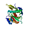
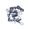
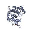
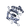
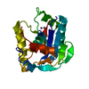
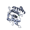
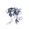
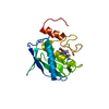
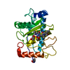
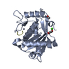
| Title | Crystal structure of the large terminase nuclease from thermophilic phage G20c with bound Cobalt | |||||||||
 Components Components | Phage terminase large subunit | |||||||||
 Keywords Keywords |  VIRAL PROTEIN / large terminase / nuclease domain / Hydrolase VIRAL PROTEIN / large terminase / nuclease domain / Hydrolase | |||||||||
| Function / homology |  Function and homology information Function and homology informationviral terminase, large subunit / viral DNA genome packaging / Hydrolases; Acting on ester bonds; Endodeoxyribonucleases producing 5'-phosphomonoesters / chromosome organization / Hydrolases; Acting on acid anhydrides; Acting on acid anhydrides to facilitate cellular and subcellular movement / endonuclease activity / ATP hydrolysis activity / ATP binding / metal ion bindingSimilarity search - Function | |||||||||
| Biological species |   Thermus phage G20c (virus) Thermus phage G20c (virus) | |||||||||
| Method | X-RAY DIFFRACTION / SYNCHROTRON / SAD / Resolution: 1.6 Å | |||||||||
 Authors Authors | Xu, R.G. / Jenkins, H.T. / Chechik, M. / Blagova, E.V. / Greive, S.J. / Antson, A.A. | |||||||||
| Funding support |  United Kingdom, 2items United Kingdom, 2items
| |||||||||
 Citation Citation | Journal: Nucleic Acids Res. / Year: 2017 Title: Viral genome packaging terminase cleaves DNA using the canonical RuvC-like two-metal catalysis mechanism. Authors: Xu, R.G. / Jenkins, H.T. / Chechik, M. / Blagova, E.V. / Lopatina, A. / Klimuk, E. / Minakhin, L. / Severinov, K. / Greive, S.J. / Antson, A.A. | |||||||||
| History |
|
- Structure visualization
| Structure viewer | Molecule: MolmilJmol/JSmol |
|---|
- Downloads & links
-Download
| PDBx/mmCIF format | 5m1o.cif.gz | 90.3 KB | Display | PDBx/mmCIF format |
|---|---|---|---|---|
| PDB format | pdb5m1o.ent.gz | 71.2 KB | Display | PDB format |
| PDBx/mmJSON format | 5m1o.json.gz | Tree view | PDBx/mmJSON format | |
| Others |  Other downloads Other downloads |
-Validation report
| Arichive directory | https://data.pdbj.org/pub/pdb/validation_reports/m1/5m1oftp://data.pdbj.org/pub/pdb/validation_reports/m1/5m1o | HTTPS FTP |
|---|
-Related structure data


-Links
 PDBj
PDBj
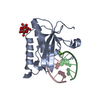
- Assembly
| Deposited unit | 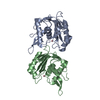
| ||||||||
|---|---|---|---|---|---|---|---|---|---|
| 1 | 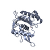
| ||||||||
| 2 | 
| ||||||||
| Unit cell |
|
-Components
| #1: Protein | Mass: 21174.803 Da / Num. of mol.: 2 Source method: isolated from a genetically manipulated source Source: (gene. exp.) Thermus phage G20c (virus) / Production host:  Escherichia coli (E. coli) / References: UniProt: A0A1L4BKS3*PLUS Escherichia coli (E. coli) / References: UniProt: A0A1L4BKS3*PLUS#2: Chemical |   Mass: 58.933 Da / Num. of mol.: 2 / Source method: obtained synthetically / Formula: Co Mass: 58.933 Da / Num. of mol.: 2 / Source method: obtained synthetically / Formula: Co#3: Water | ChemComp-HOH / | Water Mass: 18.015 Da / Num. of mol.: 319 / Source method: isolated from a natural source / Formula: H2O Mass: 18.015 Da / Num. of mol.: 319 / Source method: isolated from a natural source / Formula: H2O |
|---|
-Experimental details
-Experiment
| Experiment | Method: X-RAY DIFFRACTION / Number of used crystals: 1 |
|---|
- Sample preparation
| Crystal | Density Matthews: 1.92 Å3/Da / Density % sol: 36.1 % |
|---|---|
| Crystal grow | Temperature: 293 K / Method: vapor diffusion, sitting drop Details: 0.2 M Ammonium Tartrate, 0.1 M Bis-tris pH 5.5-6.5, 20% PEG 3350 PH range: 5.5-6.5 |
-Data collection
| Diffraction | Mean temperature: 100 K | ||||||||||||||||||
|---|---|---|---|---|---|---|---|---|---|---|---|---|---|---|---|---|---|---|---|
| Diffraction source | Source: SYNCHROTRON / Site: Diamond / Beamline: I02 / Wavelength: 0.9796 Å | ||||||||||||||||||
| Detector | Type: DECTRIS PILATUS 6M-F / Detector: PIXEL / Date: Mar 6, 2016 | ||||||||||||||||||
| Radiation | Protocol: SINGLE WAVELENGTH / Monochromatic (M) / Laue (L): M / Scattering type: x-ray | ||||||||||||||||||
| Radiation wavelength | Wavelength: 0.9796 Å / Relative weight: 1 | ||||||||||||||||||
| Reflection | Resolution: 1.6→42.92 Å / Num. obs: 40243 / % possible obs: 95.7 % / Redundancy: 2.1 % / CC1/2: 0.997 / Rmerge(I) obs: 0.072 / Rpim(I) all: 0.057 / Rrim(I) all: 0.092 / Net I/σ(I): 6.6 / Num. measured all: 86303 | ||||||||||||||||||
| Reflection shell |
|
- Processing
| Software |
| |||||||||||||||||||||||||||||||||||||||||||||||||||||||||||||||||||||||||||
|---|---|---|---|---|---|---|---|---|---|---|---|---|---|---|---|---|---|---|---|---|---|---|---|---|---|---|---|---|---|---|---|---|---|---|---|---|---|---|---|---|---|---|---|---|---|---|---|---|---|---|---|---|---|---|---|---|---|---|---|---|---|---|---|---|---|---|---|---|---|---|---|---|---|---|---|---|
| Refinement | Method to determine structure: SAD / Resolution: 1.6→42.92 Å / Cor.coef. Fo:Fc: 0.966 / Cor.coef. Fo:Fc free: 0.948 / SU B: 2.934 / SU ML: 0.095 / Cross valid method: THROUGHOUT / σ(F): 0 / ESU R: 0.111 / ESU R Free: 0.109 Details: HYDROGENS HAVE BEEN ADDED IN THE RIDING POSITIONS U VALUES : REFINED INDIVIDUALLY
| |||||||||||||||||||||||||||||||||||||||||||||||||||||||||||||||||||||||||||
| Solvent computation | Ion probe radii: 0.8 Å / Shrinkage radii: 0.8 Å / VDW probe radii: 1.2 Å | |||||||||||||||||||||||||||||||||||||||||||||||||||||||||||||||||||||||||||
| Displacement parameters | Biso max: 58.05 Å2 / Biso mean: 22.409 Å2 / Biso min: 11.18 Å2
| |||||||||||||||||||||||||||||||||||||||||||||||||||||||||||||||||||||||||||
| Refinement step | Cycle: final / Resolution: 1.6→42.92 Å
| |||||||||||||||||||||||||||||||||||||||||||||||||||||||||||||||||||||||||||
| Refine LS restraints |
| |||||||||||||||||||||||||||||||||||||||||||||||||||||||||||||||||||||||||||
| LS refinement shell | Resolution: 1.6→1.642 Å / Total num. of bins used: 20
|
