 Movie
Movie Controller


+ Open data

- Basic information
| Entry | Database: PDB / ID: 5ltd | ||||||
|---|---|---|---|---|---|---|---|
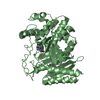
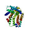
| Title | Phosphate-bound Pichia angusta Atg18 | ||||||
 Components Components | Autophagy-related protein 18 | ||||||
 Keywords Keywords | LIPID BINDING PROTEIN / membrane bound protein | ||||||
| Function / homology |  Function and homology information Function and homology informationphagophore assembly site membrane / vacuolar membrane /  autophagy / protein transport / endosome membrane autophagy / protein transport / endosome membraneSimilarity search - Function | ||||||
| Biological species |  Pichia angusta (fungus) Pichia angusta (fungus) | ||||||
| Method | X-RAY DIFFRACTION / SYNCHROTRON / MOLECULAR REPLACEMENT / Resolution: 2.5 Å | ||||||
 Authors Authors | Scacioc, A. / Kuhnel, K. | ||||||
| Funding support |  Germany, 1items Germany, 1items
| ||||||
 Citation Citation | Journal: To Be Published Title: Phopshate-bound Pichia angusta Atg18 Authors: Scacioc, A. / Kuhnel, K. | ||||||
| History |
|
- Structure visualization
| Structure viewer | Molecule: MolmilJmol/JSmol |
|---|
- Downloads & links
-Download
| PDBx/mmCIF format | 5ltd.cif.gz | 138 KB | Display | PDBx/mmCIF format |
|---|---|---|---|---|
| PDB format | pdb5ltd.ent.gz | 103.2 KB | Display | PDB format |
| PDBx/mmJSON format | 5ltd.json.gz | Tree view | PDBx/mmJSON format | |
| Others |  Other downloads Other downloads |
-Validation report
| Arichive directory | https://data.pdbj.org/pub/pdb/validation_reports/lt/5ltdftp://data.pdbj.org/pub/pdb/validation_reports/lt/5ltd | HTTPS FTP |
|---|
-Related structure data
| Related structure data |  5ltgS S: Starting model for refinement |
|---|---|
| Similar structure data |


-Links
 PDBj
PDBj- Assembly
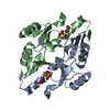
| Deposited unit | 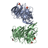
| ||||||||
|---|---|---|---|---|---|---|---|---|---|
| 1 | 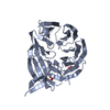
| ||||||||
| 2 | 
| ||||||||
| Unit cell |
|
-Components
| #1: Protein | Mass: 58005.082 Da / Num. of mol.: 2 Source method: isolated from a genetically manipulated source Details: two phosphate ions bound as ligands to the protein / Source: (gene. exp.) Pichia angusta (fungus) / Gene: ATG18 / Production host:  Escherichia coli (E. coli) / References: UniProt: Q5QA94 Escherichia coli (E. coli) / References: UniProt: Q5QA94#2: Chemical | ChemComp-PO4 / Phosphate  Mass: 94.971 Da / Num. of mol.: 4 / Source method: isolated from a natural source / Formula: PO4 Mass: 94.971 Da / Num. of mol.: 4 / Source method: isolated from a natural source / Formula: PO4#3: Chemical | ChemComp-EDO / | Ethylene glycol  Mass: 62.068 Da / Num. of mol.: 1 / Source method: obtained synthetically / Formula: C2H6O2 Mass: 62.068 Da / Num. of mol.: 1 / Source method: obtained synthetically / Formula: C2H6O2#4: Water | ChemComp-HOH / | Water Mass: 18.015 Da / Num. of mol.: 119 / Source method: isolated from a natural source / Formula: H2O Mass: 18.015 Da / Num. of mol.: 119 / Source method: isolated from a natural source / Formula: H2O |
|---|
-Experimental details
-Experiment
| Experiment | Method: X-RAY DIFFRACTION / Number of used crystals: 1 |
|---|
- Sample preparation
| Crystal grow | Temperature: 293 K / Method: vapor diffusion Details: 20 % (w/V) PEG 8000, 0.2 M NaCl, 0.1 M sodium phosphate dibasic-citric acid pH 4.2 |
|---|
-Data collection
| Diffraction | Mean temperature: 100 K |
|---|---|
| Diffraction source | Source: SYNCHROTRON / Site: SLS  / Beamline: X10SA / Wavelength: 0.979 Å / Beamline: X10SA / Wavelength: 0.979 Å |
| Detector | Type: PSI PILATUS 6M / Detector: PIXEL / Date: Mar 16, 2014 |
| Radiation | Protocol: SINGLE WAVELENGTH / Monochromatic (M) / Laue (L): M / Scattering type: x-ray |
| Radiation wavelength | Wavelength: 0.979 Å / Relative weight: 1 |
| Reflection | Resolution: 2.5→45 Å / Num. obs: 27376 / % possible obs: 98.3 % / Redundancy: 7.4 % / Rmerge(I) obs: 0.133 / Net I/σ(I): 12.8 |
| Reflection shell | Rmerge(I) obs: 0.645 / % possible all: 96.5 |
- Processing
| Software |
| ||||||||||||||||
|---|---|---|---|---|---|---|---|---|---|---|---|---|---|---|---|---|---|
| Refinement | Method to determine structure: MOLECULAR REPLACEMENT Starting model: 5LTG Resolution: 2.5→45 Å / Cross valid method: FREE R-VALUE
| ||||||||||||||||
| Refinement step | Cycle: LAST / Resolution: 2.5→45 Å
|
