 Movie
Movie Controller

[English] 日本語
 Yorodumi
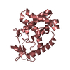
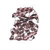
- PDB-5lm3: Plasmodium falciparum nicotinic acid mononucleotide adenylyltrans... -
+ Open data

- Basic information
| Entry | Database: PDB / ID: 5lm3 | ||||||
|---|---|---|---|---|---|---|---|
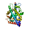
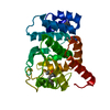
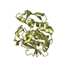
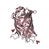
| Title | Plasmodium falciparum nicotinic acid mononucleotide adenylyltransferase complexed with APC | ||||||
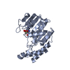
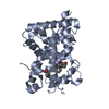
 Components Components | Nicotinate-nucleotide adenylyltransferase | ||||||
 Keywords Keywords | TRANSFERASE / Nicotinic acid mononucleotide adenylyltransferase / NaMNAT / protein crystallography / Plasmodium falciparum / drug target / malaria / NAD metabolism | ||||||
| Function / homology |  Function and homology informationnicotinamide-nucleotide adenylyltransferase / nicotinate-nucleotide adenylyltransferase activity / NAD biosynthetic process / cytoplasm Function and homology informationnicotinamide-nucleotide adenylyltransferase / nicotinate-nucleotide adenylyltransferase activity / NAD biosynthetic process / cytoplasmSimilarity search - Function | ||||||
| Biological species |  Plasmodium falciparum (malaria parasite P. falciparum) Plasmodium falciparum (malaria parasite P. falciparum) | ||||||
| Method | X-RAY DIFFRACTION / SYNCHROTRON / MOLECULAR REPLACEMENT / Resolution: 2.5 Å | ||||||
 Authors Authors | Bathke, J. / Fritz-Wolf, K. / Rahlfs, S. / Brandstaether, C. / Burkhardt, A. / Becker, K. | ||||||
| Funding support |  Germany, 1items Germany, 1items
| ||||||
 Citation Citation | Journal: J. Mol. Biol. / Year: 2016 Title: Structural and Functional Characterization of Plasmodium falciparum Nicotinic Acid Mononucleotide Adenylyltransferase. Authors: Bathke, J. / Fritz-Wolf, K. / Brandstadter, C. / Burkhardt, A. / Jortzik, E. / Rahlfs, S. / Becker, K. | ||||||
| History |
|
- Structure visualization
| Structure viewer | Molecule: MolmilJmol/JSmol |
|---|
- Downloads & links
-Download
| PDBx/mmCIF format | 5lm3.cif.gz | 57.7 KB | Display | PDBx/mmCIF format |
|---|---|---|---|---|
| PDB format | pdb5lm3.ent.gz | 40.2 KB | Display | PDB format |
| PDBx/mmJSON format | 5lm3.json.gz | Tree view | PDBx/mmJSON format | |
| Others |  Other downloads Other downloads |
-Validation report
| Arichive directory | https://data.pdbj.org/pub/pdb/validation_reports/lm/5lm3ftp://data.pdbj.org/pub/pdb/validation_reports/lm/5lm3 | HTTPS FTP |
|---|
-Related structure data
| Related structure data |  5lltSC S: Starting model for refinement C: citing same article ( |
|---|---|
| Similar structure data |


-Links
 PDBj
PDBj


- Assembly
| Deposited unit | 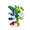
| ||||||||
|---|---|---|---|---|---|---|---|---|---|
| 1 |
| ||||||||
| Unit cell |
|
-Components
| #1: Protein | Mass: 25445.338 Da / Num. of mol.: 1 Source method: isolated from a genetically manipulated source Source: (gene. exp.) Plasmodium falciparum (isolate 3D7) (eukaryote)Strain: isolate 3D7 / Gene: PF3D7_1327600 / Production host:  Escherichia coli (E. coli) / Strain (production host): KRX Escherichia coli (E. coli) / Strain (production host): KRXReferences: UniProt: Q8IE38, nicotinate-nucleotide adenylyltransferase |
|---|---|
| #2: Chemical | ChemComp-APC /   Mass: 505.208 Da / Num. of mol.: 1 / Source method: obtained synthetically / Formula: C11H18N5O12P3 / Comment: AMP-CPP, energy-carrying molecule analogue*YM Mass: 505.208 Da / Num. of mol.: 1 / Source method: obtained synthetically / Formula: C11H18N5O12P3 / Comment: AMP-CPP, energy-carrying molecule analogue*YM |
| #3: Chemical | ChemComp-EDO / Ethylene glycol  Mass: 62.068 Da / Num. of mol.: 1 / Source method: obtained synthetically / Formula: C2H6O2 Mass: 62.068 Da / Num. of mol.: 1 / Source method: obtained synthetically / Formula: C2H6O2 |
| #4: Water | ChemComp-HOH / Water Mass: 18.015 Da / Num. of mol.: 33 / Source method: isolated from a natural source / Formula: H2O Mass: 18.015 Da / Num. of mol.: 33 / Source method: isolated from a natural source / Formula: H2O |
-Experimental details
-Experiment
| Experiment | Method: X-RAY DIFFRACTION / Number of used crystals: 1 |
|---|
- Sample preparation
| Crystal | Density Matthews: 2.43 Å3/Da / Density % sol: 49.36 % |
|---|---|
| Crystal grow | Temperature: 300 K / Method: vapor diffusion, sitting drop Details: 14 mg/ml, 0.15 M bicine, pH 8.5 and 19% PEG 6000, 5mM APC |
-Data collection
| Diffraction | Mean temperature: 100 K |
|---|---|
| Diffraction source | Source: SYNCHROTRON / Site: PETRA III, DESY / Beamline: P11 / Wavelength: 1.033208 Å |
| Detector | Type: DECTRIS PILATUS 6M / Detector: PIXEL / Date: Feb 2, 2014 |
| Radiation | Protocol: SINGLE WAVELENGTH / Monochromatic (M) / Laue (L): M / Scattering type: x-ray |
| Radiation wavelength | Wavelength: 1.033208 Å / Relative weight: 1 |
| Reflection | Resolution: 2.5→32.2 Å / Num. obs: 8537 / % possible obs: 98.2 % / Redundancy: 3.6 % / CC1/2: 0.995 / Rmerge(I) obs: 0.123 / Rsym value: 0.104 / Net I/σ(I): 8.16 |
| Reflection shell | Resolution: 2.5→2.56 Å / Redundancy: 3.7 % / Rmerge(I) obs: 0.815 / Mean I/σ(I) obs: 2.05 / CC1/2: 0.831 / % possible all: 99.5 |
- Processing
| Software |
| |||||||||||||||||||||||||||||||||||
|---|---|---|---|---|---|---|---|---|---|---|---|---|---|---|---|---|---|---|---|---|---|---|---|---|---|---|---|---|---|---|---|---|---|---|---|---|
| Refinement | Method to determine structure: MOLECULAR REPLACEMENT Starting model: 5LLT Resolution: 2.5→32.23 Å / SU ML: 0.32 / Cross valid method: FREE R-VALUE / σ(F): 1.36 / Phase error: 34.06 / Stereochemistry target values: ML
| |||||||||||||||||||||||||||||||||||
| Solvent computation | Shrinkage radii: 0.9 Å / VDW probe radii: 1.11 Å / Solvent model: FLAT BULK SOLVENT MODEL | |||||||||||||||||||||||||||||||||||
| Refinement step | Cycle: LAST / Resolution: 2.5→32.23 Å
| |||||||||||||||||||||||||||||||||||
| Refine LS restraints |
| |||||||||||||||||||||||||||||||||||
| LS refinement shell |
|
