[English] 日本語
Yorodumi
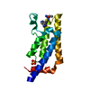
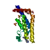
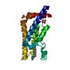
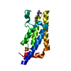
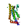
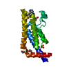
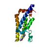
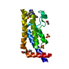
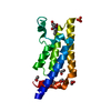
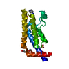
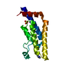
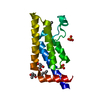
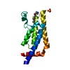
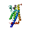
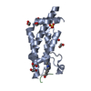
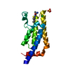
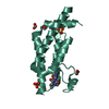
- PDB-5lj0: Crystal structure of human ATAD2 bromodomain in complex with 8-((... -

+
Open data


ID or keywords:

Loading...

-
Basic information

Entry
Database: PDB / ID: 5lj0
TitleCrystal structure of human ATAD2 bromodomain in complex with 8-(((3R,4R,5S)-3-((4,4-difluorocyclohexyl)methoxy)-5-methoxypiperidin-4-yl)amino)-3-methyl-5-(5-methylpyridin-3-yl)-1,7-naphthyridin-2(1H)-one
ComponentsATPase family AAA domain-containing protein 2
KeywordsTRANSCRIPTION / INHIBITOR / ATAD2 / BROMODOMAIN / EPIGENETICS / ATPase family AAA domain-containing protein 2
Function / homology
Function and homology information


Hydrolases; Acting on acid anhydrides; In phosphorus-containing anhydrides / TFAP2 (AP-2) family regulates transcription of growth factors and their receptors / histone binding / chromatin binding / positive regulation of DNA-templated transcription / ATP hydrolysis activity / positive regulation of transcription by RNA polymerase II / extracellular exosome / nucleoplasm / ATP binding / nucleus
Similarity search - Function
ATPase family AAA domain-containing protein ATAD2-like / AAA ATPase, AAA+ lid domain / AAA+ lid domain / ATPase, AAA-type, conserved site / AAA-protein family signature. / ATPase family associated with various cellular activities (AAA) / ATPase, AAA-type, core / Bromodomain-like / Histone Acetyltransferase; Chain A / Bromodomain ...ATPase family AAA domain-containing protein ATAD2-like / AAA ATPase, AAA+ lid domain / AAA+ lid domain / ATPase, AAA-type, conserved site / AAA-protein family signature. / ATPase family associated with various cellular activities (AAA) / ATPase, AAA-type, core / Bromodomain-like / Histone Acetyltransferase; Chain A / Bromodomain / Bromodomain profile. / bromo domain / Bromodomain / Bromodomain-like superfamily / ATPases associated with a variety of cellular activities / AAA+ ATPase domain / Up-down Bundle / P-loop containing nucleoside triphosphate hydrolase / Mainly Alpha
Similarity search - Domain/homology
Chem-6XX / ATPase family AAA domain-containing protein 2
Similarity search - Component
Biological speciesHomo sapiens (human)
MethodX-RAY DIFFRACTION / SYNCHROTRON / Resolution: 1.82 Å
AuthorsChung, C.
CitationJournal: Angew.Chem.Int.Ed.Engl. / Year: 2016
Title: A Chemical Probe for the ATAD2 Bromodomain.
Authors: Bamborough, P. / Chung, C.W. / Demont, E.H. / Furze, R.C. / Bannister, A.J. / Che, K.H. / Diallo, H. / Douault, C. / Grandi, P. / Kouzarides, T. / Michon, A.M. / Mitchell, D.J. / Prinjha, R. ...Authors: Bamborough, P. / Chung, C.W. / Demont, E.H. / Furze, R.C. / Bannister, A.J. / Che, K.H. / Diallo, H. / Douault, C. / Grandi, P. / Kouzarides, T. / Michon, A.M. / Mitchell, D.J. / Prinjha, R.K. / Rau, C. / Robson, S. / Sheppard, R.J. / Upton, R. / Watson, R.J.
History
DepositionJul 17, 2016Deposition site: PDBE / Processing site: PDBE
Revision 1.0Aug 31, 2016Provider: repository / Type: Initial release
Revision 1.1Sep 14, 2016Group: Database references

-
Structure visualization

Structure viewerMolecule:
MolmilJmol/JSmol

Downloads & links

-
Assembly

Deposited unit
A: ATPase family AAA domain-containing protein 2
hetero molecules


Theoretical massNumber of molelcules
Total (without water)16,3257
Polymers15,4541
Non-polymers8726
Water4,486249
1


  • Idetical with deposited unit
  • defined by software
TypeNameSymmetry operationNumber
identity operation1_555x,y,z1
Buried area870 Å2
ΔGint-0 kcal/mol
Surface area8170 Å2
MethodPISA
Unit cell
Length a, b, c (Å)79.158, 79.158, 135.857
Angle α, β, γ (deg.)90.00, 90.00, 120.00
Int Tables number179
Space group name H-MP6522
Components on special symmetry positions
IDModelComponents
11A-1359-

HOH

21A-1506-

HOH

-
Components

#1: Protein ATPase family AAA domain-containing protein 2 / AAA nuclear coregulator cancer-associated protein / ANCCA


Mass: 15453.514 Da / Num. of mol.: 1
Source method: isolated from a genetically manipulated source
Details: SM first two residues are residual after cleavage of the expression tag
Source: (gene. exp.) Homo sapiens (human) / Gene: ATAD2, L16, PRO2000 / Plasmid: pNIC28-Bsa4 / Production host: Escherichia coli (E. coli) / References: UniProt: Q6PL18, EC: 3.6.1.3
#2: Chemical
ChemComp-EDO / 1,2-ETHANEDIOL / ETHYLENE GLYCOL / Ethylene glycol


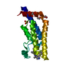
Mass: 62.068 Da / Num. of mol.: 4 / Source method: obtained synthetically / Formula: C2H6O2
#3: Chemical ChemComp-6XX / 8-(((3R,4R,5S)-3-((4,4-difluorocyclohexyl)methoxy)-5-methoxypiperidin-4-yl)amino)-3-methyl-5-(5-methylpyridin-3-yl)-1,7-naphthyridin-2(1H)-one


Mass: 527.606 Da / Num. of mol.: 1 / Source method: obtained synthetically / Formula: C28H35F2N5O3
#4: Chemical ChemComp-SO4 / SULFATE ION / Sulfate


Mass: 96.063 Da / Num. of mol.: 1 / Source method: obtained synthetically / Formula: SO4
#5: Water ChemComp-HOH / water / Water


Mass: 18.015 Da / Num. of mol.: 249 / Source method: isolated from a natural source / Formula: H2O

-
Experimental details

-
Experiment

ExperimentMethod: X-RAY DIFFRACTION / Number of used crystals: 1

-
Sample preparation

CrystalDensity Matthews: 3.98 Å3/Da / Density % sol: 69.06 %
Crystal growTemperature: 277 K / Method: vapor diffusion, sitting drop
Details: 0.1M trisHCl, pH 7.0-8.0, 1.2-1.5M ammonium sulphate, 20-25% PEG3350,277K
PH range: 7.0-8.0

-
Data collection

DiffractionMean temperature: 100 K
Diffraction sourceSource: SYNCHROTRON / Site: ESRF / Beamline: ID29 / Wavelength: 0.95373 Å
DetectorType: DECTRIS PILATUS3 6M / Detector: PIXEL / Date: Feb 7, 2014
RadiationProtocol: SINGLE WAVELENGTH / Monochromatic (M) / Laue (L): M / Scattering type: x-ray
Radiation wavelengthWavelength: 0.95373 Å / Relative weight: 1
ReflectionResolution: 1.82→135.86 Å / Num. obs: 23289 / % possible obs: 100 % / Redundancy: 10.6 % / Rmerge(I) obs: 0.072 / Net I/σ(I): 19.3
Reflection shellResolution: 1.82→1.92 Å / Redundancy: 11.1 % / Rmerge(I) obs: 0.507 / Mean I/σ(I) obs: 5.2 / % possible all: 100

-
Processing

Software
NameVersionClassification
REFMAC5.8.0073refinement
XDSdata reduction
SCALAdata scaling
RefinementResolution: 1.82→68.55 Å / Cor.coef. Fo:Fc: 0.96 / Cor.coef. Fo:Fc free: 0.958 / SU B: 3.419 / SU ML: 0.058 / Cross valid method: THROUGHOUT / ESU R: 0.095 / ESU R Free: 0.086 / Details: HYDROGENS HAVE BEEN ADDED IN THE RIDING POSITIONS
RfactorNum. reflection% reflectionSelection details
Rfree0.18256 1192 5.1 %RANDOM
Rwork0.17378 ---
obs0.17424 22036 99.97 %-
Solvent computationIon probe radii: 0.8 Å / Shrinkage radii: 0.8 Å / VDW probe radii: 1.2 Å
Displacement parametersBiso mean: 31.982 Å2
Baniso -1Baniso -2Baniso -3
1-0.02 Å20.01 Å20 Å2
2--0.02 Å2-0 Å2
3----0.07 Å2
Refinement stepCycle: LAST / Resolution: 1.82→68.55 Å
ProteinNucleic acidLigandSolventTotal
Num. atoms1084 0 59 249 1392
Refine LS restraints
Refine-IDTypeDev idealDev ideal targetNumber
X-RAY DIFFRACTIONr_bond_refined_d0.0050.0191196
X-RAY DIFFRACTIONr_bond_other_d0.0030.021164
X-RAY DIFFRACTIONr_angle_refined_deg0.9491.9871618
X-RAY DIFFRACTIONr_angle_other_deg0.74232682
X-RAY DIFFRACTIONr_dihedral_angle_1_deg4.1835137
X-RAY DIFFRACTIONr_dihedral_angle_2_deg32.50223.28164
X-RAY DIFFRACTIONr_dihedral_angle_3_deg11.01715221
X-RAY DIFFRACTIONr_dihedral_angle_4_deg12.8581515
X-RAY DIFFRACTIONr_chiral_restr0.0580.2179
X-RAY DIFFRACTIONr_gen_planes_refined0.0030.0211294
X-RAY DIFFRACTIONr_gen_planes_other0.0010.02267
X-RAY DIFFRACTIONr_nbd_refined
X-RAY DIFFRACTIONr_nbd_other
X-RAY DIFFRACTIONr_nbtor_refined
X-RAY DIFFRACTIONr_nbtor_other
X-RAY DIFFRACTIONr_xyhbond_nbd_refined
X-RAY DIFFRACTIONr_xyhbond_nbd_other
X-RAY DIFFRACTIONr_metal_ion_refined
X-RAY DIFFRACTIONr_metal_ion_other
X-RAY DIFFRACTIONr_symmetry_vdw_refined
X-RAY DIFFRACTIONr_symmetry_vdw_other
X-RAY DIFFRACTIONr_symmetry_hbond_refined
X-RAY DIFFRACTIONr_symmetry_hbond_other
X-RAY DIFFRACTIONr_symmetry_metal_ion_refined
X-RAY DIFFRACTIONr_symmetry_metal_ion_other
X-RAY DIFFRACTIONr_mcbond_it1.2942.895527
X-RAY DIFFRACTIONr_mcbond_other1.2842.885526
X-RAY DIFFRACTIONr_mcangle_it2.1566.475659
X-RAY DIFFRACTIONr_mcangle_other2.166.491660
X-RAY DIFFRACTIONr_scbond_it1.9913.426668
X-RAY DIFFRACTIONr_scbond_other1.9863.413664
X-RAY DIFFRACTIONr_scangle_it
X-RAY DIFFRACTIONr_scangle_other3.4027.398949
X-RAY DIFFRACTIONr_long_range_B_refined8.20415.821699
X-RAY DIFFRACTIONr_long_range_B_other8.20415.8191699
X-RAY DIFFRACTIONr_rigid_bond_restr
X-RAY DIFFRACTIONr_sphericity_free
X-RAY DIFFRACTIONr_sphericity_bonded
LS refinement shellResolution: 1.822→1.869 Å / Total num. of bins used: 20
RfactorNum. reflection% reflection
Rfree0.266 74 -
Rwork0.205 1601 -
obs--100 %
Refinement TLS params.Method: refined / Origin x: -12.5882 Å / Origin y: 40.853 Å / Origin z: 3.4195 Å
111213212223313233
T0.0155 Å2-0.023 Å20.0026 Å2-0.0917 Å2-0.0356 Å2--0.027 Å2
L2.3605 °2-0.1356 °2-0.6309 °2-0.0232 °20.1179 °2--0.8138 °2
S-0.0507 Å °-0.2264 Å °0.0951 Å °-0.003 Å °0.0334 Å °-0.0082 Å °0.0226 Å °0.0313 Å °0.0174 Å °

+
About Yorodumi

-
News

-
Feb 9, 2022. New format data for meta-information of EMDB entries

New format data for meta-information of EMDB entries

  • Version 3 of the EMDB header file is now the official format.
  • The previous official version 1.9 will be removed from the archive.

Related info.:EMDB header

External links:wwPDB to switch to version 3 of the EMDB data model

-
Aug 12, 2020. Covid-19 info

Covid-19 info

URL: https://pdbj.org/emnavi/covid19.php

New page: Covid-19 featured information page in EM Navigator.

Related info.:Covid-19 info / Mar 5, 2020. Novel coronavirus structure data

+
Mar 5, 2020. Novel coronavirus structure data

Novel coronavirus structure data

Related info.:Yorodumi Speices / Aug 12, 2020. Covid-19 info

External links:COVID-19 featured content - PDBj / Molecule of the Month (242):Coronavirus Proteases

+
Jan 31, 2019. EMDB accession codes are about to change! (news from PDBe EMDB page)

EMDB accession codes are about to change! (news from PDBe EMDB page)

  • The allocation of 4 digits for EMDB accession codes will soon come to an end. Whilst these codes will remain in use, new EMDB accession codes will include an additional digit and will expand incrementally as the available range of codes is exhausted. The current 4-digit format prefixed with “EMD-” (i.e. EMD-XXXX) will advance to a 5-digit format (i.e. EMD-XXXXX), and so on. It is currently estimated that the 4-digit codes will be depleted around Spring 2019, at which point the 5-digit format will come into force.
  • The EM Navigator/Yorodumi systems omit the EMD- prefix.

Related info.:Q: What is EMD? / ID/Accession-code notation in Yorodumi/EM Navigator

External links:EMDB Accession Codes are Changing Soon! / Contact to PDBj

+
Jul 12, 2017. Major update of PDB

Major update of PDB

  • wwPDB released updated PDB data conforming to the new PDBx/mmCIF dictionary.
  • This is a major update changing the version number from 4 to 5, and with Remediation, in which all the entries are updated.
  • In this update, many items about electron microscopy experimental information are reorganized (e.g. em_software).
  • Now, EM Navigator and Yorodumi are based on the updated data.

External links:wwPDB Remediation / Enriched Model Files Conforming to OneDep Data Standards Now Available in the PDB FTP Archive

-
Yorodumi

Thousand views of thousand structures

  • Yorodumi is a browser for structure data from EMDB, PDB, SASBDB, etc.
  • This page is also the successor to EM Navigator detail page, and also detail information page/front-end page for Omokage search.
  • The word "yorodu" (or yorozu) is an old Japanese word meaning "ten thousand". "mi" (miru) is to see.

Related info.:EMDB / PDB / SASBDB / Comparison of 3 databanks / Yorodumi Search / Aug 31, 2016. New EM Navigator & Yorodumi / Yorodumi Papers / Jmol/JSmol / Function and homology information / Changes in new EM Navigator and Yorodumi

Read more