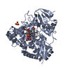
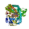
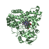
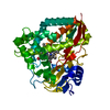
Entry Database : PDB / ID : 5k5xTitle Crystal structure of human PDGFRA Platelet-derived growth factor receptor alpha Keywords / / Function / homology Function Domain/homology Component
/ / / / / / / / / / / / / / / / / / / / / / / / / / / / / / / / / / / / / / / / / / / / / / / / / / / / / / / / / / / / / / / / / / / / / / / / / / / / / / / / / / / / / / / / / / / / / / / / / / / / / / / / / / / / / / / / / / / / / / / / / / / / / / / / / Biological species Homo sapiens (human)Method / / / Resolution : 2.168 Å Authors Yan, X.E. / Liang, L. / Yun, C.H. Funding support Organization Grant number Country
Journal : Biochem.Biophys.Res.Commun. / Year : 2016Title : Structural and biochemical studies of the PDGFRA kinase domainAuthors : Liang, L. / Yan, X.E. / Yin, Y. / Yun, C.H. History Deposition May 24, 2016 Deposition site / Processing site Revision 1.0 Aug 17, 2016 Provider / Type Revision 1.1 Oct 4, 2017 Group / Database references / Derived calculationsCategory / diffrn_detector / pdbx_struct_oper_listItem / _diffrn_detector.detector / _pdbx_struct_oper_list.symmetry_operationRevision 1.2 Nov 8, 2023 Group / Database references / Refinement descriptionCategory chem_comp_atom / chem_comp_bond ... chem_comp_atom / chem_comp_bond / database_2 / pdbx_initial_refinement_model Item / _database_2.pdbx_database_accession
Show all Show less
 Movie
Movie Controller


 Open data
 Basic information
 Components
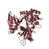
 PDGFRA
  Keywords
 Function and homology information
 Authors
 China, 1items
  Citation
 Structure visualization
 Downloads & links
 Other downloads


 PDBj
PDBj


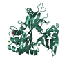
 Assembly


 Mass: 96.063 Da / Num. of mol.: 3 / Source method: obtained synthetically / Formula: SO4
 Mass: 18.015 Da / Num. of mol.: 148 / Source method: isolated from a natural source / Formula: H2O
 Sample preparation
 Processing