 Movie
Movie Controller

[English] 日本語
 Yorodumi
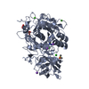
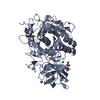
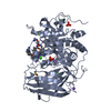
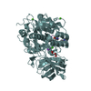
- PDB-5jxi: Structure of the unliganded form of the proprotein convertase fur... -
+ Open data

- Basic information
| Entry | Database: PDB / ID: 5jxi | |||||||||
|---|---|---|---|---|---|---|---|---|---|---|
| Title | Structure of the unliganded form of the proprotein convertase furin in presence of EDTA. | |||||||||
 Components Components | Furin | |||||||||
 Keywords Keywords | HYDROLASE / protease / apo-structure / proteolysis | |||||||||
| Function / homology |  Function and homology informationfurin / nerve growth factor production / dibasic protein processing / plasma lipoprotein particle remodeling / NGF processing / Assembly of active LPL and LIPC lipase complexes / peptide biosynthetic process / negative regulation of transforming growth factor beta1 production / signal peptide processing / regulation of cholesterol transport ...furin / nerve growth factor production / dibasic protein processing / plasma lipoprotein particle remodeling / NGF processing / Assembly of active LPL and LIPC lipase complexes / peptide biosynthetic process / negative regulation of transforming growth factor beta1 production / signal peptide processing / regulation of cholesterol transport / negative regulation of low-density lipoprotein particle receptor catabolic process / cytokine precursor processing / Pre-NOTCH Processing in Golgi / secretion by cell / Synthesis and processing of ENV and VPU / nerve growth factor binding / Formation of the cornified envelope / Signaling by PDGF / trans-Golgi network transport vesicle / blastocyst formation / Elastic fibre formation / heparan sulfate binding / Signaling by NODAL / positive regulation of membrane protein ectodomain proteolysis / zymogen activation / regulation of endopeptidase activity / peptide hormone processing / CD163 mediating an anti-inflammatory response / regulation of protein catabolic process / Activation of Matrix Metalloproteinases / TGF-beta receptor signaling activates SMADs / protein maturation / Uptake and function of anthrax toxins / Collagen degradation / collagen catabolic process / extracellular matrix disassembly / regulation of signal transduction / Removal of aminoterminal propeptides from gamma-carboxylated proteins / negative regulation of inflammatory response to antigenic stimulus / viral life cycle / serine-type peptidase activity / extracellular matrix organization / transforming growth factor beta receptor signaling pathway / peptide binding / SMAD2/SMAD3:SMAD4 heterotrimer regulates transcription / trans-Golgi network / serine-type endopeptidase inhibitor activity / protein processing / Golgi lumen / heparin binding / peptidase activity / viral translation / endopeptidase activity / Induction of Cell-Cell Fusion / protease binding / amyloid fibril formation / Potential therapeutics for SARS / Attachment and Entry / positive regulation of viral entry into host cell / viral protein processing / endosome membrane / membrane raft / Amyloid fiber formation / Golgi membrane / serine-type endopeptidase activity / cell surface / endoplasmic reticulum / extracellular exosome / extracellular region / membrane / metal ion binding / plasma membrane Function and homology informationfurin / nerve growth factor production / dibasic protein processing / plasma lipoprotein particle remodeling / NGF processing / Assembly of active LPL and LIPC lipase complexes / peptide biosynthetic process / negative regulation of transforming growth factor beta1 production / signal peptide processing / regulation of cholesterol transport ...furin / nerve growth factor production / dibasic protein processing / plasma lipoprotein particle remodeling / NGF processing / Assembly of active LPL and LIPC lipase complexes / peptide biosynthetic process / negative regulation of transforming growth factor beta1 production / signal peptide processing / regulation of cholesterol transport / negative regulation of low-density lipoprotein particle receptor catabolic process / cytokine precursor processing / Pre-NOTCH Processing in Golgi / secretion by cell / Synthesis and processing of ENV and VPU / nerve growth factor binding / Formation of the cornified envelope / Signaling by PDGF / trans-Golgi network transport vesicle / blastocyst formation / Elastic fibre formation / heparan sulfate binding / Signaling by NODAL / positive regulation of membrane protein ectodomain proteolysis / zymogen activation / regulation of endopeptidase activity / peptide hormone processing / CD163 mediating an anti-inflammatory response / regulation of protein catabolic process / Activation of Matrix Metalloproteinases / TGF-beta receptor signaling activates SMADs / protein maturation / Uptake and function of anthrax toxins / Collagen degradation / collagen catabolic process / extracellular matrix disassembly / regulation of signal transduction / Removal of aminoterminal propeptides from gamma-carboxylated proteins / negative regulation of inflammatory response to antigenic stimulus / viral life cycle / serine-type peptidase activity / extracellular matrix organization / transforming growth factor beta receptor signaling pathway / peptide binding / SMAD2/SMAD3:SMAD4 heterotrimer regulates transcription / trans-Golgi network / serine-type endopeptidase inhibitor activity / protein processing / Golgi lumen / heparin binding / peptidase activity / viral translation / endopeptidase activity / Induction of Cell-Cell Fusion / protease binding / amyloid fibril formation / Potential therapeutics for SARS / Attachment and Entry / positive regulation of viral entry into host cell / viral protein processing / endosome membrane / membrane raft / Amyloid fiber formation / Golgi membrane / serine-type endopeptidase activity / cell surface / endoplasmic reticulum / extracellular exosome / extracellular region / membrane / metal ion binding / plasma membraneSimilarity search - Function | |||||||||
| Biological species |  Homo sapiens (human) Homo sapiens (human) | |||||||||
| Method | X-RAY DIFFRACTION / SYNCHROTRON / MOLECULAR REPLACEMENT / Resolution: 2 Å | |||||||||
 Authors Authors | Dahms, S.O. / Arciniega, M. / Steinmetzer, T. / Huber, R. / Than, M.E. | |||||||||
 Citation Citation | Journal: Proc.Natl.Acad.Sci.USA / Year: 2016 Title: Structure of the unliganded form of the proprotein convertase furin suggests activation by a substrate-induced mechanism. Authors: Dahms, S.O. / Arciniega, M. / Steinmetzer, T. / Huber, R. / Than, M.E. | |||||||||
| History |
|
- Structure visualization
| Structure viewer | Molecule: MolmilJmol/JSmol |
|---|
- Downloads & links
-Download
| PDBx/mmCIF format | 5jxi.cif.gz | 121.6 KB | Display | PDBx/mmCIF format |
|---|---|---|---|---|
| PDB format | pdb5jxi.ent.gz | 90.3 KB | Display | PDB format |
| PDBx/mmJSON format | 5jxi.json.gz | Tree view | PDBx/mmJSON format | |
| Others |  Other downloads Other downloads |
-Validation report
| Arichive directory | https://data.pdbj.org/pub/pdb/validation_reports/jx/5jxiftp://data.pdbj.org/pub/pdb/validation_reports/jx/5jxi | HTTPS FTP |
|---|
-Related structure data
| Related structure data |  5jxgC  5jxhC  5jxjC  4rydS S: Starting model for refinement C: citing same article ( |
|---|---|
| Similar structure data |


-Links
 PDBj
PDBj


- Assembly
| Deposited unit | 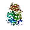
| |||||||||
|---|---|---|---|---|---|---|---|---|---|---|
| 1 |
| |||||||||
| Unit cell |
| |||||||||
| Components on special symmetry positions |
|
-Components
| #1: Protein | / Dibasic-processing enzyme / Paired basic amino acid residue-cleaving enzyme / PACE Mass: 52388.602 Da / Num. of mol.: 1 Source method: isolated from a genetically manipulated source Source: (gene. exp.) Homo sapiens (human) / Gene: FURIN, FUR, PACE, PCSK3 / Cell line (production host): HEK293S / Production host: Homo sapiens (human) / References: UniProt: P09958, furin | ||||
|---|---|---|---|---|---|
| #2: Chemical | ChemComp-CA /   Mass: 40.078 Da / Num. of mol.: 1 / Source method: obtained synthetically / Formula: Ca Mass: 40.078 Da / Num. of mol.: 1 / Source method: obtained synthetically / Formula: Ca | ||||
| #3: Chemical | ChemComp-NA /   Mass: 22.990 Da / Num. of mol.: 7 / Source method: obtained synthetically / Formula: Na Mass: 22.990 Da / Num. of mol.: 7 / Source method: obtained synthetically / Formula: Na#4: Chemical | ChemComp-CL / | Chloride  Mass: 35.453 Da / Num. of mol.: 1 / Source method: obtained synthetically / Formula: Cl Mass: 35.453 Da / Num. of mol.: 1 / Source method: obtained synthetically / Formula: Cl#5: Water | ChemComp-HOH / | Water Mass: 18.015 Da / Num. of mol.: 520 / Source method: isolated from a natural source / Formula: H2O Mass: 18.015 Da / Num. of mol.: 520 / Source method: isolated from a natural source / Formula: H2O |
-Experimental details
-Experiment
| Experiment | Method: X-RAY DIFFRACTION / Number of used crystals: 1 |
|---|
- Sample preparation
| Crystal | Density Matthews: 3.76 Å3/Da / Density % sol: 67.3 % |
|---|---|
| Crystal grow | Temperature: 293.15 K / Method: vapor diffusion, sitting drop Details: crystallization solution: 100 mM MES, 200 mM K/NaH2PO4, pH 5.5-6.0, 3-4 M NaCl, 3% DMSO; reservoir solution: 3-4 M NaCl |
-Data collection
| Diffraction | Mean temperature: 100 K |
|---|---|
| Diffraction source | Source: SYNCHROTRON / Site: BESSY  / Beamline: 14.1 / Wavelength: 0.918409 Å / Beamline: 14.1 / Wavelength: 0.918409 Å |
| Detector | Type: DECTRIS PILATUS3 6M / Detector: PIXEL / Date: Nov 24, 2014 |
| Radiation | Protocol: SINGLE WAVELENGTH / Monochromatic (M) / Laue (L): M / Scattering type: x-ray |
| Radiation wavelength | Wavelength: 0.918409 Å / Relative weight: 1 |
| Reflection | Resolution: 2→46.2 Å / Num. obs: 54336 / % possible obs: 98.9 % / Redundancy: 4.9 % / Rsym value: 0.116 / Net I/σ(I): 14.3 |
| Reflection shell | Resolution: 2→2.12 Å / Rsym value: 0.595 |
- Processing
| Software |
| ||||||||||||||||||||||||||||||||||||||||||||||||||||||||||||||||||||||||||||||||||||||||||||||||||||||||||||||||||||||||||||||||||||||||||||
|---|---|---|---|---|---|---|---|---|---|---|---|---|---|---|---|---|---|---|---|---|---|---|---|---|---|---|---|---|---|---|---|---|---|---|---|---|---|---|---|---|---|---|---|---|---|---|---|---|---|---|---|---|---|---|---|---|---|---|---|---|---|---|---|---|---|---|---|---|---|---|---|---|---|---|---|---|---|---|---|---|---|---|---|---|---|---|---|---|---|---|---|---|---|---|---|---|---|---|---|---|---|---|---|---|---|---|---|---|---|---|---|---|---|---|---|---|---|---|---|---|---|---|---|---|---|---|---|---|---|---|---|---|---|---|---|---|---|---|---|---|---|
| Refinement | Method to determine structure: MOLECULAR REPLACEMENT Starting model: 4RYD Resolution: 2→43.354 Å / SU ML: 0.17 / Cross valid method: FREE R-VALUE / σ(F): 1.36 / Phase error: 17.03
| ||||||||||||||||||||||||||||||||||||||||||||||||||||||||||||||||||||||||||||||||||||||||||||||||||||||||||||||||||||||||||||||||||||||||||||
| Solvent computation | Shrinkage radii: 0.9 Å / VDW probe radii: 1.11 Å | ||||||||||||||||||||||||||||||||||||||||||||||||||||||||||||||||||||||||||||||||||||||||||||||||||||||||||||||||||||||||||||||||||||||||||||
| Refinement step | Cycle: LAST / Resolution: 2→43.354 Å
| ||||||||||||||||||||||||||||||||||||||||||||||||||||||||||||||||||||||||||||||||||||||||||||||||||||||||||||||||||||||||||||||||||||||||||||
| Refine LS restraints |
| ||||||||||||||||||||||||||||||||||||||||||||||||||||||||||||||||||||||||||||||||||||||||||||||||||||||||||||||||||||||||||||||||||||||||||||
| LS refinement shell |
|
