 Movie
Movie Controller

[English] 日本語
 Yorodumi
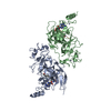
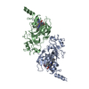
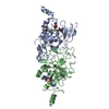
- PDB-5jj0: Structure of G9a SET-domain with Histone H3K9M peptide and excess SAH -
+ Open data

- Basic information
| Entry | Database: PDB / ID: 5jj0 | ||||||
|---|---|---|---|---|---|---|---|
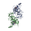
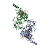
| Title | Structure of G9a SET-domain with Histone H3K9M peptide and excess SAH | ||||||
 Components Components |
| ||||||
 Keywords Keywords |  TRANSFERASE / SET-domain / Histone methyl transferase / histone peptide TRANSFERASE / SET-domain / Histone methyl transferase / histone peptide | ||||||
| Function / homology |  Function and homology information Function and homology informationregulation of protein modification process / histone H3K56 methyltransferase activity / : / phenotypic switching / neuron fate specification / : / [histone H3]-lysine9 N-methyltransferase / histone H3K27 methyltransferase activity / histone H3K9 methyltransferase activity / histone H3K9me2 methyltransferase activity ...regulation of protein modification process / histone H3K56 methyltransferase activity / : / phenotypic switching / neuron fate specification / : / [histone H3]-lysine9 N-methyltransferase / histone H3K27 methyltransferase activity / histone H3K9 methyltransferase activity / histone H3K9me2 methyltransferase activity / peptidyl-lysine dimethylation / synaptonemal complex assembly / negative regulation of autophagosome assembly / protein-lysine N-methyltransferase activity / oocyte development / C2H2 zinc finger domain binding / fertilization / : / cellular response to cocaine / : / organ growth / Transcriptional Regulation by E2F6 / spermatid development / behavioral response to cocaine / regulation of DNA replication / RNA Polymerase I Transcription Initiation / Transcriptional Regulation by VENTX / long-term memory / Chromatin modifying enzymes / epigenetic regulation of gene expression / response to fungicide / Transferases; Transferring one-carbon groups; Methyltransferases / cellular response to starvation / telomere organization / RNA Polymerase I Promoter Opening / Interleukin-7 signaling / Assembly of the ORC complex at the origin of replication / DNA methylation / Condensation of Prophase Chromosomes / transcription corepressor binding / HCMV Late Events / Chromatin modifications during the maternal to zygotic transition (MZT) / ERCC6 (CSB) and EHMT2 (G9a) positively regulate rRNA expression / SIRT1 negatively regulates rRNA expression / PRC2 methylates histones and DNA / Defective pyroptosis / promoter-specific chromatin binding / HDACs deacetylate histones / RNA Polymerase I Promoter Escape / RNA polymerase II transcription regulatory region sequence-specific DNA binding / Transcriptional regulation by small RNAs / Formation of the beta-catenin:TCF transactivating complex / RUNX1 regulates genes involved in megakaryocyte differentiation and platelet function / Activated PKN1 stimulates transcription of AR (androgen receptor) regulated genes KLK2 and KLK3 / NoRC negatively regulates rRNA expression / B-WICH complex positively regulates rRNA expression / HDMs demethylate histones / Regulation of TP53 Activity through Methylation / PKMTs methylate histone lysines / RMTs methylate histone arginines / Meiotic recombination / Pre-NOTCH Transcription and Translation / nucleosome assembly / Activation of anterior HOX genes in hindbrain development during early embryogenesis / HCMV Early Events / Transcriptional regulation of granulopoiesis / structural constituent of chromatin / nucleosome / cellular response to xenobiotic stimulus / p53 binding / gene expression / RUNX1 regulates transcription of genes involved in differentiation of HSCs / chromatin organization / Factors involved in megakaryocyte development and platelet production / HATs acetylate histones / Senescence-Associated Secretory Phenotype (SASP) / response to ethanol / Oxidative Stress Induced Senescence / Estrogen-dependent gene expression / nuclear speck / cadherin binding / Amyloid fiber formation / protein heterodimerization activity / chromatin / negative regulation of transcription by RNA polymerase II / protein-containing complex / DNA binding / extracellular exosome / zinc ion binding / extracellular region / nucleoplasm / membrane / nucleusSimilarity search - Function | ||||||
| Biological species |  Homo sapiens (human) Homo sapiens (human) | ||||||
| Method | X-RAY DIFFRACTION / SYNCHROTRON / MOLECULAR REPLACEMENT / Resolution: 1.72 Å | ||||||
 Authors Authors | Jayaram, H. / Bellon, S.F. / Poy, F. | ||||||
 Citation Citation | Journal: Proc.Natl.Acad.Sci.USA / Year: 2016 Title: S-adenosyl methionine is necessary for inhibition of the methyltransferase G9a by the lysine 9 to methionine mutation on histone H3. Authors: Jayaram, H. / Hoelper, D. / Jain, S.U. / Cantone, N. / Lundgren, S.M. / Poy, F. / Allis, C.D. / Cummings, R. / Bellon, S. / Lewis, P.W. | ||||||
| History |
|
- Structure visualization
| Structure viewer | Molecule: MolmilJmol/JSmol |
|---|
- Downloads & links
-Download
| PDBx/mmCIF format | 5jj0.cif.gz | 132 KB | Display | PDBx/mmCIF format |
|---|---|---|---|---|
| PDB format | pdb5jj0.ent.gz | 101 KB | Display | PDB format |
| PDBx/mmJSON format | 5jj0.json.gz | Tree view | PDBx/mmJSON format | |
| Others |  Other downloads Other downloads |
-Validation report
| Arichive directory | https://data.pdbj.org/pub/pdb/validation_reports/jj/5jj0ftp://data.pdbj.org/pub/pdb/validation_reports/jj/5jj0 | HTTPS FTP |
|---|
-Related structure data
| Related structure data |  5jhnC  5jinC  5jiyC 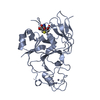 2o8jS C: citing same article ( S: Starting model for refinement |
|---|---|
| Similar structure data |

-Links
 PDBj
PDBj


- Assembly
| Deposited unit | 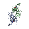
| ||||||||
|---|---|---|---|---|---|---|---|---|---|
| 1 |
| ||||||||
| Unit cell |
|
-Components
| #1: Protein | Mass: 31661.904 Da / Num. of mol.: 2 Fragment: SET domain of Histone-lysine N-methyltransferase EHMT2 G9a, UNP residues 882-1155 Source method: isolated from a genetically manipulated source Source: (gene. exp.) Homo sapiens (human) / Gene: EHMT2, BAT8, C6orf30, G9A, KMT1C, NG36 / Production host:  Escherichia coli (E. coli) Escherichia coli (E. coli)References: UniProt: Q96KQ7, Transferases; Transferring one-carbon groups; Methyltransferases, histone-lysine N-methyltransferase#2: Protein/peptide | Mass: 1139.284 Da / Num. of mol.: 2 Source method: isolated from a genetically manipulated source Source: (gene. exp.) Homo sapiens (human) / Production host: Escherichia coli (E. coli) / References: UniProt: P68431*PLUS#3: Chemical | ChemComp-ZN /   Mass: 65.409 Da / Num. of mol.: 8 / Source method: obtained synthetically / Formula: Zn Mass: 65.409 Da / Num. of mol.: 8 / Source method: obtained synthetically / Formula: Zn#4: Chemical | S-Adenosyl methionine  Mass: 398.437 Da / Num. of mol.: 2 / Source method: obtained synthetically / Formula: C15H22N6O5S Mass: 398.437 Da / Num. of mol.: 2 / Source method: obtained synthetically / Formula: C15H22N6O5S#5: Water | ChemComp-HOH / | Water Mass: 18.015 Da / Num. of mol.: 130 / Source method: isolated from a natural source / Formula: H2O Mass: 18.015 Da / Num. of mol.: 130 / Source method: isolated from a natural source / Formula: H2O |
|---|
-Experimental details
-Experiment
| Experiment | Method: X-RAY DIFFRACTION / Number of used crystals: 1 |
|---|
- Sample preparation
| Crystal | Density Matthews: 2.47 Å3/Da / Density % sol: 50.22 % |
|---|---|
| Crystal grow | Temperature: 298 K / Method: vapor diffusion, sitting drop / pH: 7 Details: 0.2 M Tri-ammonium citrate pH 7 and 20% w/v polyethylene glycol 3350 |
-Data collection
| Diffraction | Mean temperature: 178 K |
|---|---|
| Diffraction source | Source: SYNCHROTRON / Site: APS  / Beamline: 14-BM-C / Wavelength: 1.001 Å / Beamline: 14-BM-C / Wavelength: 1.001 Å |
| Detector | Type: ADSC QUANTUM 210 / Detector: CCD / Date: Jan 1, 2013 |
| Radiation | Protocol: SINGLE WAVELENGTH / Monochromatic (M) / Laue (L): M / Scattering type: x-ray |
| Radiation wavelength | Wavelength: 1.001 Å / Relative weight: 1 |
| Reflection | Resolution: 1.72→45.91 Å / Num. obs: 63270 / % possible obs: 98.73 % / Redundancy: 3.6 % / Net I/σ(I): 10.13 |
| Reflection shell | Resolution: 1.72→1.79 Å / Redundancy: 3.7 % / Rmerge(I) obs: 0.703 / Mean I/σ(I) obs: 2.2 / % possible all: 87.49 |
- Processing
| Software |
| ||||||||||||||||||||||||||||||||||||||||||||||||||||||||||||||||||||||||||||||||||||||||||||||||||||||||||||||||||||||||||||||||||||||||||||||||||||||||||||||||||||||||||||||||||||||
|---|---|---|---|---|---|---|---|---|---|---|---|---|---|---|---|---|---|---|---|---|---|---|---|---|---|---|---|---|---|---|---|---|---|---|---|---|---|---|---|---|---|---|---|---|---|---|---|---|---|---|---|---|---|---|---|---|---|---|---|---|---|---|---|---|---|---|---|---|---|---|---|---|---|---|---|---|---|---|---|---|---|---|---|---|---|---|---|---|---|---|---|---|---|---|---|---|---|---|---|---|---|---|---|---|---|---|---|---|---|---|---|---|---|---|---|---|---|---|---|---|---|---|---|---|---|---|---|---|---|---|---|---|---|---|---|---|---|---|---|---|---|---|---|---|---|---|---|---|---|---|---|---|---|---|---|---|---|---|---|---|---|---|---|---|---|---|---|---|---|---|---|---|---|---|---|---|---|---|---|---|---|---|---|
| Refinement | Method to determine structure: MOLECULAR REPLACEMENT Starting model: 2O8J Resolution: 1.72→45.91 Å / Cor.coef. Fo:Fc: 0.949 / Cor.coef. Fo:Fc free: 0.93 / SU B: 3.309 / SU ML: 0.105 / Cross valid method: THROUGHOUT / ESU R: 0.125 / ESU R Free: 0.121 / Details: HYDROGENS HAVE BEEN ADDED IN THE RIDING POSITIONS
| ||||||||||||||||||||||||||||||||||||||||||||||||||||||||||||||||||||||||||||||||||||||||||||||||||||||||||||||||||||||||||||||||||||||||||||||||||||||||||||||||||||||||||||||||||||||
| Solvent computation | Ion probe radii: 0.8 Å / Shrinkage radii: 0.8 Å / VDW probe radii: 1.2 Å | ||||||||||||||||||||||||||||||||||||||||||||||||||||||||||||||||||||||||||||||||||||||||||||||||||||||||||||||||||||||||||||||||||||||||||||||||||||||||||||||||||||||||||||||||||||||
| Displacement parameters | Biso mean: 31.777 Å2
| ||||||||||||||||||||||||||||||||||||||||||||||||||||||||||||||||||||||||||||||||||||||||||||||||||||||||||||||||||||||||||||||||||||||||||||||||||||||||||||||||||||||||||||||||||||||
| Refinement step | Cycle: 1 / Resolution: 1.72→45.91 Å
| ||||||||||||||||||||||||||||||||||||||||||||||||||||||||||||||||||||||||||||||||||||||||||||||||||||||||||||||||||||||||||||||||||||||||||||||||||||||||||||||||||||||||||||||||||||||
| Refine LS restraints |
|