 Movie
Movie Controller


+ Open data

- Basic information
| Entry | Database: PDB / ID: 5jgk | ||||||
|---|---|---|---|---|---|---|---|
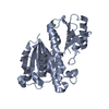
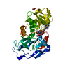
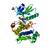
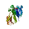
| Title | Crystal structure of GtmA in complex with SAH | ||||||
 Components Components | UbiE/COQ5 family methyltransferase, putative | ||||||
 Keywords Keywords |  TRANSFERASE / Aspergillus fumigatus / Methyltransferase / S-ADENOSYL-L-HOMOCYSTEINE / Gliotoxin / Resistance TRANSFERASE / Aspergillus fumigatus / Methyltransferase / S-ADENOSYL-L-HOMOCYSTEINE / Gliotoxin / Resistance | ||||||
| Function / homology | ubiE/COQ5 methyltransferase family / S-adenosylmethionine-dependent methyltransferase activity / methylation / S-adenosyl-L-methionine-dependent methyltransferase superfamily / S-ADENOSYL-L-HOMOCYSTEINE / UbiE/COQ5 family methyltransferase, putative Function and homology information Function and homology information | ||||||
| Biological species |  Aspergillus fumigatus Z5 (mold) Aspergillus fumigatus Z5 (mold) | ||||||
| Method | X-RAY DIFFRACTION / SYNCHROTRON / MOLECULAR REPLACEMENT / Resolution: 1.33 Å | ||||||
 Authors Authors | Dolan, S.K. / Bock, T. / Hering, V. / Jones, G.W. / Blankenfeldt, W. / Dolye, S. | ||||||
 Citation Citation | Journal: Open Biol / Year: 2017 Title: Structural, mechanistic and functional insight into gliotoxinbis-thiomethylation inAspergillus fumigatus. Authors: Dolan, S.K. / Bock, T. / Hering, V. / Owens, R.A. / Jones, G.W. / Blankenfeldt, W. / Doyle, S. | ||||||
| History |
|
- Structure visualization
| Structure viewer | Molecule: MolmilJmol/JSmol |
|---|
- Downloads & links
-Download
| PDBx/mmCIF format | 5jgk.cif.gz | 333.3 KB | Display | PDBx/mmCIF format |
|---|---|---|---|---|
| PDB format | pdb5jgk.ent.gz | 286.2 KB | Display | PDB format |
| PDBx/mmJSON format | 5jgk.json.gz | Tree view | PDBx/mmJSON format | |
| Others |  Other downloads Other downloads |
-Validation report
| Arichive directory | https://data.pdbj.org/pub/pdb/validation_reports/jg/5jgkftp://data.pdbj.org/pub/pdb/validation_reports/jg/5jgk | HTTPS FTP |
|---|
-Related structure data


-Links
 PDBj
PDBj
- Assembly
| Deposited unit | 
| ||||||||
|---|---|---|---|---|---|---|---|---|---|
| 1 | 
| ||||||||
| 2 | 
| ||||||||
| Unit cell |
|
-Components
| #1: Protein | Mass: 32455.912 Da / Num. of mol.: 2 Source method: isolated from a genetically manipulated source Source: (gene. exp.) Aspergillus fumigatus Z5 (mold) / Gene: Y699_02735 / Plasmid: pET19m / Production host:  Escherichia coli (E. coli) / References: UniProt: A0A0J5Q3C4 Escherichia coli (E. coli) / References: UniProt: A0A0J5Q3C4#2: Chemical | S-Adenosyl-L-homocysteine  Type: L-peptide linking / Mass: 384.411 Da / Num. of mol.: 2 / Source method: obtained synthetically / Formula: C14H20N6O5S Type: L-peptide linking / Mass: 384.411 Da / Num. of mol.: 2 / Source method: obtained synthetically / Formula: C14H20N6O5S#3: Chemical | ChemComp-SO4 / Sulfate  Mass: 96.063 Da / Num. of mol.: 5 / Source method: obtained synthetically / Formula: SO4 Mass: 96.063 Da / Num. of mol.: 5 / Source method: obtained synthetically / Formula: SO4#4: Water | ChemComp-HOH / | Water Mass: 18.015 Da / Num. of mol.: 401 / Source method: isolated from a natural source / Formula: H2O Mass: 18.015 Da / Num. of mol.: 401 / Source method: isolated from a natural source / Formula: H2O |
|---|
-Experimental details
-Experiment
| Experiment | Method: X-RAY DIFFRACTION / Number of used crystals: 1 |
|---|
- Sample preparation
| Crystal | Density Matthews: 2.27 Å3/Da / Density % sol: 45.9 % |
|---|---|
| Crystal grow | Temperature: 277 K / Method: vapor diffusion, sitting drop Details: 2.4 M ammonium sulfate, 0.2 M sodium chloride, 0.1 M MES pH 6.0 |
-Data collection
| Diffraction | Mean temperature: 100 K | |||||||||||||||
|---|---|---|---|---|---|---|---|---|---|---|---|---|---|---|---|---|
| Diffraction source | Source: SYNCHROTRON / Site: ESRF  / Beamline: ID30B / Wavelength: 0.9762 Å / Beamline: ID30B / Wavelength: 0.9762 Å | |||||||||||||||
| Detector | Type: DECTRIS PILATUS 6M-F / Detector: PIXEL / Date: Dec 14, 2015 | |||||||||||||||
| Radiation | Protocol: SINGLE WAVELENGTH / Monochromatic (M) / Laue (L): M / Scattering type: x-ray | |||||||||||||||
| Radiation wavelength | Wavelength: 0.9762 Å / Relative weight: 1 | |||||||||||||||
| Reflection | Resolution: 1.33→45.12 Å / Num. obs: 130776 / % possible obs: 98.7 % / Redundancy: 6.5 % / Biso Wilson estimate: 14.79 Å2 / CC1/2: 0.999 / Rmerge(I) obs: 0.064 / Rpim(I) all: 0.027 / Rrim(I) all: 0.07 / Net I/σ(I): 14.4 / Num. measured all: 852355 / Scaling rejects: 196 | |||||||||||||||
| Reflection shell |
|
- Processing
| Software |
| |||||||||||||||||||||||||||||||||||||||||||||||||||||||||||||||||||||||||||||||||||||||||||||||||||||||||||||||||||||||||||||||||||||||||||||||||||||||||||||||||||||||||||||||||||||||||||||||||||||||||||||||||||||||||
|---|---|---|---|---|---|---|---|---|---|---|---|---|---|---|---|---|---|---|---|---|---|---|---|---|---|---|---|---|---|---|---|---|---|---|---|---|---|---|---|---|---|---|---|---|---|---|---|---|---|---|---|---|---|---|---|---|---|---|---|---|---|---|---|---|---|---|---|---|---|---|---|---|---|---|---|---|---|---|---|---|---|---|---|---|---|---|---|---|---|---|---|---|---|---|---|---|---|---|---|---|---|---|---|---|---|---|---|---|---|---|---|---|---|---|---|---|---|---|---|---|---|---|---|---|---|---|---|---|---|---|---|---|---|---|---|---|---|---|---|---|---|---|---|---|---|---|---|---|---|---|---|---|---|---|---|---|---|---|---|---|---|---|---|---|---|---|---|---|---|---|---|---|---|---|---|---|---|---|---|---|---|---|---|---|---|---|---|---|---|---|---|---|---|---|---|---|---|---|---|---|---|---|---|---|---|---|---|---|---|---|---|---|---|---|---|---|---|---|
| Refinement | Method to determine structure: MOLECULAR REPLACEMENT / Resolution: 1.33→45.117 Å / SU ML: 0.1 / Cross valid method: FREE R-VALUE / σ(F): 1.34 / Phase error: 15.58
| |||||||||||||||||||||||||||||||||||||||||||||||||||||||||||||||||||||||||||||||||||||||||||||||||||||||||||||||||||||||||||||||||||||||||||||||||||||||||||||||||||||||||||||||||||||||||||||||||||||||||||||||||||||||||
| Solvent computation | Shrinkage radii: 0.9 Å / VDW probe radii: 1.11 Å | |||||||||||||||||||||||||||||||||||||||||||||||||||||||||||||||||||||||||||||||||||||||||||||||||||||||||||||||||||||||||||||||||||||||||||||||||||||||||||||||||||||||||||||||||||||||||||||||||||||||||||||||||||||||||
| Displacement parameters | Biso max: 63.31 Å2 / Biso mean: 22.3843 Å2 / Biso min: 9.06 Å2 | |||||||||||||||||||||||||||||||||||||||||||||||||||||||||||||||||||||||||||||||||||||||||||||||||||||||||||||||||||||||||||||||||||||||||||||||||||||||||||||||||||||||||||||||||||||||||||||||||||||||||||||||||||||||||
| Refinement step | Cycle: final / Resolution: 1.33→45.117 Å
| |||||||||||||||||||||||||||||||||||||||||||||||||||||||||||||||||||||||||||||||||||||||||||||||||||||||||||||||||||||||||||||||||||||||||||||||||||||||||||||||||||||||||||||||||||||||||||||||||||||||||||||||||||||||||
| Refine LS restraints |
| |||||||||||||||||||||||||||||||||||||||||||||||||||||||||||||||||||||||||||||||||||||||||||||||||||||||||||||||||||||||||||||||||||||||||||||||||||||||||||||||||||||||||||||||||||||||||||||||||||||||||||||||||||||||||
| LS refinement shell | Refine-ID: X-RAY DIFFRACTION / Total num. of bins used: 30
|
