 Movie
Movie Controller

[English] 日本語
 Yorodumi
- PDB-5i8w: Crystal structure of B. pseudomallei FabI in complex with NAD and... -
+ Open data

- Basic information
| Entry | Database: PDB / ID: 5i8w | ||||||
|---|---|---|---|---|---|---|---|
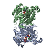
| Title | Crystal structure of B. pseudomallei FabI in complex with NAD and PT401 | ||||||
 Components Components | Enoyl-[acyl-carrier-protein] reductase [NADH] | ||||||
 Keywords Keywords |  OXIDOREDUCTASE / enoyl-ACP reductase / Burkholderia pseudomallei / diphenyl ethers / slow-binding inhibitors OXIDOREDUCTASE / enoyl-ACP reductase / Burkholderia pseudomallei / diphenyl ethers / slow-binding inhibitors | ||||||
| Function / homology |  Function and homology information Function and homology informationenoyl-[acyl-carrier-protein] reductase (NADH) / enoyl-[acyl-carrier-protein] reductase (NADH) activity / fatty acid biosynthetic process / nucleotide bindingSimilarity search - Function | ||||||
| Biological species |  Burkholderia pseudomallei (bacteria) Burkholderia pseudomallei (bacteria) | ||||||
| Method | X-RAY DIFFRACTION / MOLECULAR REPLACEMENT / Resolution: 1.629 Å | ||||||
 Authors Authors | Hirschbeck, M.W. / Eltschkner, S. / Tonge, P.J. / Kisker, C. | ||||||
| Funding support |  Germany, 1items Germany, 1items
| ||||||
 Citation Citation | Journal: Biochemistry / Year: 2017 Title: Rationalizing the Binding Kinetics for the Inhibition of the Burkholderia pseudomallei FabI1 Enoyl-ACP Reductase. Authors: Neckles, C. / Eltschkner, S. / Cummings, J.E. / Hirschbeck, M. / Daryaee, F. / Bommineni, G.R. / Zhang, Z. / Spagnuolo, L. / Yu, W. / Davoodi, S. / Slayden, R.A. / Kisker, C. / Tonge, P.J. | ||||||
| History |
|
- Structure visualization
| Structure viewer | Molecule: MolmilJmol/JSmol |
|---|
- Downloads & links
-Download
| PDBx/mmCIF format | 5i8w.cif.gz | 118.6 KB | Display | PDBx/mmCIF format |
|---|---|---|---|---|
| PDB format | pdb5i8w.ent.gz | 90.9 KB | Display | PDB format |
| PDBx/mmJSON format | 5i8w.json.gz | Tree view | PDBx/mmJSON format | |
| Others |  Other downloads Other downloads |
-Validation report
| Arichive directory | https://data.pdbj.org/pub/pdb/validation_reports/i8/5i8wftp://data.pdbj.org/pub/pdb/validation_reports/i8/5i8w | HTTPS FTP |
|---|
-Related structure data
| Related structure data |  5i7eC  5i7fC  5i7sC  5i7vC  5i8zC  5i9lC  5i9mC  5i9nC  5iflC  3ek2S S: Starting model for refinement C: citing same article ( |
|---|---|
| Similar structure data |


-Links
 PDBj
PDBj

- Assembly
| Deposited unit | 
| ||||||||||||
|---|---|---|---|---|---|---|---|---|---|---|---|---|---|
| 1 | 
| ||||||||||||
| Unit cell |
| ||||||||||||
| Components on special symmetry positions |
|
-Components
| #1: Protein | Mass: 29325.391 Da / Num. of mol.: 1 Source method: isolated from a genetically manipulated source Source: (gene. exp.) Burkholderia pseudomallei (bacteria)Gene: fabI, fabI_2, ACT79_25590, AM256_11615, AM257_11635, AMS56_08605, DP46_2957, DP49_6839, ERS012314_03793, ERS012372_03823, SY87_14645, TR70_1075, VU09_13745 Production host: Escherichia coli (E. coli)References: UniProt: A0A069B9A4, UniProt: A0A0H3HP34*PLUS, enoyl-[acyl-carrier-protein] reductase (NADH) |
|---|---|
| #2: Chemical | ChemComp-NAD / Nicotinamide adenine dinucleotide  Mass: 663.425 Da / Num. of mol.: 1 / Source method: obtained synthetically / Formula: C21H27N7O14P2 / Comment: NAD*YM Mass: 663.425 Da / Num. of mol.: 1 / Source method: obtained synthetically / Formula: C21H27N7O14P2 / Comment: NAD*YM |
| #3: Chemical | ChemComp-69H /   Mass: 302.383 Da / Num. of mol.: 1 / Source method: obtained synthetically / Formula: C19H23FO2 Mass: 302.383 Da / Num. of mol.: 1 / Source method: obtained synthetically / Formula: C19H23FO2 |
| #4: Water | ChemComp-HOH / Water Mass: 18.015 Da / Num. of mol.: 262 / Source method: isolated from a natural source / Formula: H2O Mass: 18.015 Da / Num. of mol.: 262 / Source method: isolated from a natural source / Formula: H2O |
-Experimental details
-Experiment
| Experiment | Method: X-RAY DIFFRACTION / Number of used crystals: 1 |
|---|
- Sample preparation
| Crystal | Density Matthews: 2.06 Å3/Da / Density % sol: 40.35 % |
|---|---|
| Crystal grow | Temperature: 293 K / Method: vapor diffusion, hanging drop / Details: 0.1 M Bistris, pH 6.5, 36 % PEG 300 |
-Data collection
| Diffraction | Mean temperature: 100 K |
|---|---|
| Diffraction source | Source: ROTATING ANODE / Type: RIGAKU MICROMAX-007 HF / Wavelength: 1.542 Å |
| Detector | Type: RIGAKU RAXIS HTC / Detector: IMAGE PLATE / Date: Jan 11, 2013 |
| Radiation | Protocol: SINGLE WAVELENGTH / Monochromatic (M) / Laue (L): M / Scattering type: x-ray |
| Radiation wavelength | Wavelength: 1.542 Å / Relative weight: 1 |
| Reflection | Resolution: 1.629→28.74 Å / Num. obs: 29346 / % possible obs: 93 % / Redundancy: 4.1 % / Rmerge(I) obs: 0.063 / Net I/σ(I): 15.9 |
| Reflection shell | Resolution: 1.63→1.72 Å / Redundancy: 4.1 % / Rmerge(I) obs: 0.119 / Mean I/σ(I) obs: 8.7 / % possible all: 88.4 |
- Processing
| Software |
| ||||||||||||||||||||||||||||||||||||||||||||||||||||||||||||||||||||||||||||||||||||||||||||||||||||||||||||||||||||||||||||||||||||||||||||||||||||||||||||||||||||||||||||||||||||||||||||||||||||||||
|---|---|---|---|---|---|---|---|---|---|---|---|---|---|---|---|---|---|---|---|---|---|---|---|---|---|---|---|---|---|---|---|---|---|---|---|---|---|---|---|---|---|---|---|---|---|---|---|---|---|---|---|---|---|---|---|---|---|---|---|---|---|---|---|---|---|---|---|---|---|---|---|---|---|---|---|---|---|---|---|---|---|---|---|---|---|---|---|---|---|---|---|---|---|---|---|---|---|---|---|---|---|---|---|---|---|---|---|---|---|---|---|---|---|---|---|---|---|---|---|---|---|---|---|---|---|---|---|---|---|---|---|---|---|---|---|---|---|---|---|---|---|---|---|---|---|---|---|---|---|---|---|---|---|---|---|---|---|---|---|---|---|---|---|---|---|---|---|---|---|---|---|---|---|---|---|---|---|---|---|---|---|---|---|---|---|---|---|---|---|---|---|---|---|---|---|---|---|---|---|---|---|
| Refinement | Method to determine structure: MOLECULAR REPLACEMENT Starting model: 3EK2 Resolution: 1.629→26.584 Å / SU ML: 0.13 / Cross valid method: FREE R-VALUE / σ(F): 1.34 / Phase error: 19.51
| ||||||||||||||||||||||||||||||||||||||||||||||||||||||||||||||||||||||||||||||||||||||||||||||||||||||||||||||||||||||||||||||||||||||||||||||||||||||||||||||||||||||||||||||||||||||||||||||||||||||||
| Solvent computation | Shrinkage radii: 0.9 Å / VDW probe radii: 1.11 Å | ||||||||||||||||||||||||||||||||||||||||||||||||||||||||||||||||||||||||||||||||||||||||||||||||||||||||||||||||||||||||||||||||||||||||||||||||||||||||||||||||||||||||||||||||||||||||||||||||||||||||
| Refinement step | Cycle: LAST / Resolution: 1.629→26.584 Å
| ||||||||||||||||||||||||||||||||||||||||||||||||||||||||||||||||||||||||||||||||||||||||||||||||||||||||||||||||||||||||||||||||||||||||||||||||||||||||||||||||||||||||||||||||||||||||||||||||||||||||
| Refine LS restraints |
| ||||||||||||||||||||||||||||||||||||||||||||||||||||||||||||||||||||||||||||||||||||||||||||||||||||||||||||||||||||||||||||||||||||||||||||||||||||||||||||||||||||||||||||||||||||||||||||||||||||||||
| LS refinement shell |
| ||||||||||||||||||||||||||||||||||||||||||||||||||||||||||||||||||||||||||||||||||||||||||||||||||||||||||||||||||||||||||||||||||||||||||||||||||||||||||||||||||||||||||||||||||||||||||||||||||||||||
| Refinement TLS params. | Method: refined / Refine-ID: X-RAY DIFFRACTION
| ||||||||||||||||||||||||||||||||||||||||||||||||||||||||||||||||||||||||||||||||||||||||||||||||||||||||||||||||||||||||||||||||||||||||||||||||||||||||||||||||||||||||||||||||||||||||||||||||||||||||
| Refinement TLS group |
|
