 Movie
Movie Controller


+ Open data

- Basic information
| Entry | Database: PDB / ID: 5hww | ||||||
|---|---|---|---|---|---|---|---|
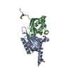
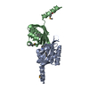
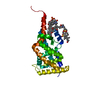
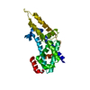
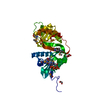
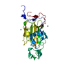
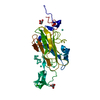
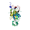
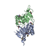
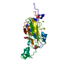
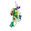
| Title | Crystal structure of PAS1 complexed with 1,2,4-TMB | ||||||
 Components Components | Sensor histidine kinase TodS | ||||||
 Keywords Keywords |  TRANSFERASE / PAS / Two-component signal transduction / toluene TRANSFERASE / PAS / Two-component signal transduction / toluene | ||||||
| Function / homology |  Function and homology informationhistidine kinase / phosphorelay sensor kinase activity / ATP binding / cytoplasm Function and homology informationhistidine kinase / phosphorelay sensor kinase activity / ATP binding / cytoplasmSimilarity search - Function | ||||||
| Biological species |  Pseudomonas putida (bacteria) Pseudomonas putida (bacteria) | ||||||
| Method | X-RAY DIFFRACTION / SYNCHROTRON / Resolution: 2 Å | ||||||
 Authors Authors | Hwang, J. / Koh, S. | ||||||
 Citation Citation | Journal: J. Biol. Chem. / Year: 2016 Title: Molecular Insights into Toluene Sensing in the TodS/TodT Signal Transduction System. Authors: Koh, S. / Hwang, J. / Guchhait, K. / Lee, E.G. / Kim, S.Y. / Kim, S. / Lee, S. / Chung, J.M. / Jung, H.S. / Lee, S.J. / Ryu, C.M. / Lee, S.G. / Oh, T.K. / Kwon, O. / Kim, M.H. | ||||||
| History |
|
- Structure visualization
| Structure viewer | Molecule: MolmilJmol/JSmol |
|---|
- Downloads & links
-Download
| PDBx/mmCIF format | 5hww.cif.gz | 109.5 KB | Display | PDBx/mmCIF format |
|---|---|---|---|---|
| PDB format | pdb5hww.ent.gz | 83.9 KB | Display | PDB format |
| PDBx/mmJSON format | 5hww.json.gz | Tree view | PDBx/mmJSON format | |
| Others |  Other downloads Other downloads |
-Validation report
| Arichive directory | https://data.pdbj.org/pub/pdb/validation_reports/hw/5hwwftp://data.pdbj.org/pub/pdb/validation_reports/hw/5hww | HTTPS FTP |
|---|
-Related structure data

-Links
 PDBj
PDBj


- Assembly
| Deposited unit | 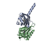
| ||||||||
|---|---|---|---|---|---|---|---|---|---|
| 1 |
| ||||||||
| Unit cell |
|
-Components
| #1: Protein | Mass: 14353.434 Da / Num. of mol.: 2 / Fragment: PAS1 (UNP RESIDUES 43-168) Source method: isolated from a genetically manipulated source Source: (gene. exp.) Pseudomonas putida (strain F1 / ATCC 700007) (bacteria)Strain: F1 / ATCC 700007 / Gene: todS, Pput_2872 / Production host: Escherichia coli (E. coli) / References: UniProt: A5W4E3, histidine kinase#2: Chemical | 1,2,4-Trimethylbenzene  Mass: 120.192 Da / Num. of mol.: 2 / Source method: obtained synthetically / Formula: C9H12 Mass: 120.192 Da / Num. of mol.: 2 / Source method: obtained synthetically / Formula: C9H12#3: Water | ChemComp-HOH / | Water Mass: 18.015 Da / Num. of mol.: 154 / Source method: isolated from a natural source / Formula: H2O Mass: 18.015 Da / Num. of mol.: 154 / Source method: isolated from a natural source / Formula: H2O |
|---|
-Experimental details
-Experiment
| Experiment | Method: X-RAY DIFFRACTION |
|---|
- Sample preparation
| Crystal | Density Matthews: 2.03 Å3/Da / Density % sol: 39.44 % |
|---|---|
| Crystal grow | Temperature: 294.15 K / Method: vapor diffusion, sitting drop / pH: 8 / Details: 3.0 M ammonium phosphate, 0.1 M Tris-HCl |
-Data collection
| Diffraction | Mean temperature: 173.15 K |
|---|---|
| Diffraction source | Source: SYNCHROTRON / Site: PAL/PLS  / Beamline: 5C (4A) / Wavelength: 0.9795 Å / Beamline: 5C (4A) / Wavelength: 0.9795 Å |
| Detector | Type: ADSC QUANTUM 315 / Detector: CCD / Date: Jul 6, 2012 |
| Radiation | Protocol: SINGLE WAVELENGTH / Monochromatic (M) / Laue (L): M / Scattering type: x-ray |
| Radiation wavelength | Wavelength: 0.9795 Å / Relative weight: 1 |
| Reflection | Resolution: 1.96→50 Å / Num. obs: 17149 / % possible obs: 98.8 % / Redundancy: 13.7 % / Net I/σ(I): 49.98 |
- Processing
| Software |
| ||||||||||||||||||||||||||||||||||||||||||||||||||||||||||||||||||||||||||||||||||||||||||||||||||||||||||||||||||||||||||||||||||||||||||||||||||||||||||||||||||||||||||||||||||||||
|---|---|---|---|---|---|---|---|---|---|---|---|---|---|---|---|---|---|---|---|---|---|---|---|---|---|---|---|---|---|---|---|---|---|---|---|---|---|---|---|---|---|---|---|---|---|---|---|---|---|---|---|---|---|---|---|---|---|---|---|---|---|---|---|---|---|---|---|---|---|---|---|---|---|---|---|---|---|---|---|---|---|---|---|---|---|---|---|---|---|---|---|---|---|---|---|---|---|---|---|---|---|---|---|---|---|---|---|---|---|---|---|---|---|---|---|---|---|---|---|---|---|---|---|---|---|---|---|---|---|---|---|---|---|---|---|---|---|---|---|---|---|---|---|---|---|---|---|---|---|---|---|---|---|---|---|---|---|---|---|---|---|---|---|---|---|---|---|---|---|---|---|---|---|---|---|---|---|---|---|---|---|---|---|
| Refinement | Resolution: 2→30 Å / Cor.coef. Fo:Fc: 0.952 / Cor.coef. Fo:Fc free: 0.907 / SU B: 9.857 / SU ML: 0.135 / Cross valid method: THROUGHOUT / ESU R: 0.226 / ESU R Free: 0.21 / Stereochemistry target values: MAXIMUM LIKELIHOOD
| ||||||||||||||||||||||||||||||||||||||||||||||||||||||||||||||||||||||||||||||||||||||||||||||||||||||||||||||||||||||||||||||||||||||||||||||||||||||||||||||||||||||||||||||||||||||
| Solvent computation | Ion probe radii: 0.8 Å / Shrinkage radii: 0.8 Å / VDW probe radii: 1.2 Å / Solvent model: MASK | ||||||||||||||||||||||||||||||||||||||||||||||||||||||||||||||||||||||||||||||||||||||||||||||||||||||||||||||||||||||||||||||||||||||||||||||||||||||||||||||||||||||||||||||||||||||
| Displacement parameters | Biso mean: 29.175 Å2
| ||||||||||||||||||||||||||||||||||||||||||||||||||||||||||||||||||||||||||||||||||||||||||||||||||||||||||||||||||||||||||||||||||||||||||||||||||||||||||||||||||||||||||||||||||||||
| Refinement step | Cycle: 1 / Resolution: 2→30 Å
| ||||||||||||||||||||||||||||||||||||||||||||||||||||||||||||||||||||||||||||||||||||||||||||||||||||||||||||||||||||||||||||||||||||||||||||||||||||||||||||||||||||||||||||||||||||||
| Refine LS restraints |
|