 Movie
Movie Controller


+ Open data

- Basic information
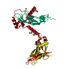
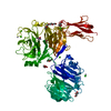
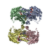
| Entry | Database: PDB / ID: 5hlz | ||||||
|---|---|---|---|---|---|---|---|
| Title | Structure of Pro-Activin A Complex at 2.85 A resolution | ||||||
 Components Components | (Inhibin beta A chain) x 2 | ||||||
 Keywords Keywords |  SIGNALING PROTEIN / Growth factor / Precursor / Signalling SIGNALING PROTEIN / Growth factor / Precursor / Signalling | ||||||
| Function / homology |  Function and homology informationactivin A complex / inhibin A complex / cardiac fibroblast cell development / regulation of follicle-stimulating hormone secretion / negative regulation of B cell differentiation / positive regulation of ovulation / GABAergic neuron differentiation / Antagonism of Activin by Follistatin / negative regulation of follicle-stimulating hormone secretion / progesterone secretion ...activin A complex / inhibin A complex / cardiac fibroblast cell development / regulation of follicle-stimulating hormone secretion / negative regulation of B cell differentiation / positive regulation of ovulation / GABAergic neuron differentiation / Antagonism of Activin by Follistatin / negative regulation of follicle-stimulating hormone secretion / progesterone secretion / type II activin receptor binding / striatal medium spiny neuron differentiation / negative regulation of macrophage differentiation / Glycoprotein hormones / positive regulation of follicle-stimulating hormone secretion / cellular response to oxygen-glucose deprivation / hemoglobin biosynthetic process / cellular response to follicle-stimulating hormone stimulus / cellular response to cholesterol / Signaling by BMP / negative regulation of phosphorylation / activin receptor signaling pathway / Signaling by Activin / mesodermal cell differentiation / positive regulation of extrinsic apoptotic signaling pathway in absence of ligand / SMAD protein signal transduction / cellular response to angiotensin / positive regulation of transcription by RNA polymerase III / odontogenesis / response to aldosterone / negative regulation of G1/S transition of mitotic cell cycle / endodermal cell differentiation / roof of mouth development / eyelid development in camera-type eye / peptide hormone binding / positive regulation of SMAD protein signal transduction / negative regulation of type II interferon production / hair follicle development / positive regulation of collagen biosynthetic process / hematopoietic progenitor cell differentiation / extrinsic apoptotic signaling pathway / ovarian follicle development / positive regulation of protein metabolic process / erythrocyte differentiation / positive regulation of erythrocyte differentiation / cytokine activity / growth factor activity / defense response / hormone activity / negative regulation of cell growth / autophagy / cytokine-mediated signaling pathway / male gonad development / cell-cell signaling / nervous system development / cellular response to hypoxia / transcription by RNA polymerase II / cell differentiation / positive regulation of ERK1 and ERK2 cascade / cell surface receptor signaling pathway / positive regulation of protein phosphorylation / negative regulation of cell population proliferation / protein-containing complex binding / positive regulation of gene expression / regulation of transcription by RNA polymerase II / perinuclear region of cytoplasm / positive regulation of DNA-templated transcription / positive regulation of transcription by RNA polymerase II / extracellular space / extracellular region / identical protein binding Function and homology informationactivin A complex / inhibin A complex / cardiac fibroblast cell development / regulation of follicle-stimulating hormone secretion / negative regulation of B cell differentiation / positive regulation of ovulation / GABAergic neuron differentiation / Antagonism of Activin by Follistatin / negative regulation of follicle-stimulating hormone secretion / progesterone secretion ...activin A complex / inhibin A complex / cardiac fibroblast cell development / regulation of follicle-stimulating hormone secretion / negative regulation of B cell differentiation / positive regulation of ovulation / GABAergic neuron differentiation / Antagonism of Activin by Follistatin / negative regulation of follicle-stimulating hormone secretion / progesterone secretion / type II activin receptor binding / striatal medium spiny neuron differentiation / negative regulation of macrophage differentiation / Glycoprotein hormones / positive regulation of follicle-stimulating hormone secretion / cellular response to oxygen-glucose deprivation / hemoglobin biosynthetic process / cellular response to follicle-stimulating hormone stimulus / cellular response to cholesterol / Signaling by BMP / negative regulation of phosphorylation / activin receptor signaling pathway / Signaling by Activin / mesodermal cell differentiation / positive regulation of extrinsic apoptotic signaling pathway in absence of ligand / SMAD protein signal transduction / cellular response to angiotensin / positive regulation of transcription by RNA polymerase III / odontogenesis / response to aldosterone / negative regulation of G1/S transition of mitotic cell cycle / endodermal cell differentiation / roof of mouth development / eyelid development in camera-type eye / peptide hormone binding / positive regulation of SMAD protein signal transduction / negative regulation of type II interferon production / hair follicle development / positive regulation of collagen biosynthetic process / hematopoietic progenitor cell differentiation / extrinsic apoptotic signaling pathway / ovarian follicle development / positive regulation of protein metabolic process / erythrocyte differentiation / positive regulation of erythrocyte differentiation / cytokine activity / growth factor activity / defense response / hormone activity / negative regulation of cell growth / autophagy / cytokine-mediated signaling pathway / male gonad development / cell-cell signaling / nervous system development / cellular response to hypoxia / transcription by RNA polymerase II / cell differentiation / positive regulation of ERK1 and ERK2 cascade / cell surface receptor signaling pathway / positive regulation of protein phosphorylation / negative regulation of cell population proliferation / protein-containing complex binding / positive regulation of gene expression / regulation of transcription by RNA polymerase II / perinuclear region of cytoplasm / positive regulation of DNA-templated transcription / positive regulation of transcription by RNA polymerase II / extracellular space / extracellular region / identical protein bindingSimilarity search - Function | ||||||
| Biological species |  Homo sapiens (human) Homo sapiens (human) | ||||||
| Method | X-RAY DIFFRACTION / SYNCHROTRON / MOLECULAR REPLACEMENT / Resolution: 2.851 Å | ||||||
 Authors Authors | Wang, X. / Fischer, G. / Hyvonen, M. | ||||||
 Citation Citation | Journal: Nat Commun / Year: 2016 Title: Structure and activation of pro-activin A. Authors: Wang, X. / Fischer, G. / Hyvonen, M. | ||||||
| History |
|
- Structure visualization
| Structure viewer | Molecule: MolmilJmol/JSmol |
|---|
- Downloads & links
-Download
| PDBx/mmCIF format | 5hlz.cif.gz | 236.5 KB | Display | PDBx/mmCIF format |
|---|---|---|---|---|
| PDB format | pdb5hlz.ent.gz | 196.2 KB | Display | PDB format |
| PDBx/mmJSON format | 5hlz.json.gz | Tree view | PDBx/mmJSON format | |
| Others |  Other downloads Other downloads |
-Validation report
| Arichive directory | https://data.pdbj.org/pub/pdb/validation_reports/hl/5hlzftp://data.pdbj.org/pub/pdb/validation_reports/hl/5hlz | HTTPS FTP |
|---|
-Related structure data


-Links
 PDBj
PDBj


- Assembly
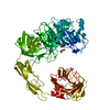
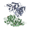
| Deposited unit | 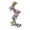
| ||||||||
|---|---|---|---|---|---|---|---|---|---|
| 1 | 
| ||||||||
| 2 | 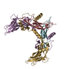
| ||||||||
| Unit cell |
|
-Components
| #1: Protein | Mass: 30272.439 Da / Num. of mol.: 4 / Fragment: Pro domain, UNP Residues 30-305 Mutation: C35S, C38S, deletion of K259-D282, furin cleavage site (RRRRR) replaced by HRV 3C protease cleavage site (LEVLFQGP),C35S, C38S, deletion of K259-D282, furin cleavage site replaced by HRV 3C ...Mutation: C35S, C38S, deletion of K259-D282, furin cleavage site (RRRRR) replaced by HRV 3C protease cleavage site (LEVLFQGP),C35S, C38S, deletion of K259-D282, furin cleavage site replaced by HRV 3C protease cleavage site (LEVLFQGP),C35S, C38S, deletion of K259-D282, furin cleavage site (RRRRR) replaced by HRV 3C protease cleavage site (LEVLFQGP),C35S, C38S, deletion of K259-D282, furin cleavage site replaced by HRV 3C protease cleavage site (LEVLFQGP) Source method: isolated from a genetically manipulated source Source: (gene. exp.) Homo sapiens (human) / Gene: INHBA / Details (production host): N-terminal His6-tag / Production host:  Escherichia coli BL21(DE3) (bacteria) / References: UniProt: P08476 Escherichia coli BL21(DE3) (bacteria) / References: UniProt: P08476#2: Protein | Mass: 12991.865 Da / Num. of mol.: 4 / Fragment: Mature domain, UNP Residues 311-426 Source method: isolated from a genetically manipulated source Source: (gene. exp.) Homo sapiens (human) / Gene: INHBA / Production host: Escherichia coli BL21(DE3) (bacteria) / References: UniProt: P08476#3: Water | ChemComp-HOH / | Water Mass: 18.015 Da / Num. of mol.: 10 / Source method: isolated from a natural source / Formula: H2O Mass: 18.015 Da / Num. of mol.: 10 / Source method: isolated from a natural source / Formula: H2O |
|---|
-Experimental details
-Experiment
| Experiment | Method: X-RAY DIFFRACTION / Number of used crystals: 1 |
|---|
- Sample preparation
| Crystal | Density Matthews: 1.67 Å3/Da / Density % sol: 26.24 % / Description: Rods |
|---|---|
| Crystal grow | Temperature: 292 K / Method: vapor diffusion, sitting drop / pH: 8 Details: 20% w/v polyethylene glycol 3350, 0.2 M calcium chloride; cryo: 15% v/v PEG 400 added |
-Data collection
| Diffraction | Mean temperature: 100 K |
|---|---|
| Diffraction source | Source: SYNCHROTRON / Site: Diamond  / Beamline: I04 / Wavelength: 0.97949 Å / Beamline: I04 / Wavelength: 0.97949 Å |
| Detector | Type: DECTRIS PILATUS3 6M / Detector: PIXEL / Date: Dec 15, 2014 / Details: Mirrors |
| Radiation | Monochromator: Si(111) / Protocol: SINGLE WAVELENGTH / Monochromatic (M) / Laue (L): M / Scattering type: x-ray |
| Radiation wavelength | Wavelength: 0.97949 Å / Relative weight: 1 |
| Reflection | Resolution: 2.85→44.54 Å / Num. obs: 26028 / % possible obs: 99.9 % / Observed criterion σ(I): -3 / Redundancy: 5.1 % / Biso Wilson estimate: 36 Å2 / CC1/2: 0.997 / Rmerge(I) obs: 0.114 / Rsym value: 0.141 / Net I/σ(I): 9.7 |
| Reflection shell | Resolution: 2.85→3.01 Å / Redundancy: 4.9 % / Rmerge(I) obs: 0.948 / Mean I/σ(I) obs: 1.6 / % possible all: 99.8 |
- Processing
| Software |
| |||||||||||||||||||||||||||||||||||||||||||||||||||||||||||||||||||||||||||||
|---|---|---|---|---|---|---|---|---|---|---|---|---|---|---|---|---|---|---|---|---|---|---|---|---|---|---|---|---|---|---|---|---|---|---|---|---|---|---|---|---|---|---|---|---|---|---|---|---|---|---|---|---|---|---|---|---|---|---|---|---|---|---|---|---|---|---|---|---|---|---|---|---|---|---|---|---|---|---|
| Refinement | Method to determine structure: MOLECULAR REPLACEMENT Starting model: Structure of the uncleaved growth factor Resolution: 2.851→40.9 Å / SU ML: 0.48 / Cross valid method: THROUGHOUT / σ(F): 1.96 / Phase error: 32.41 / Details: NCS restraint was included in the refinement.
| |||||||||||||||||||||||||||||||||||||||||||||||||||||||||||||||||||||||||||||
| Solvent computation | Shrinkage radii: 0.9 Å / VDW probe radii: 1.11 Å | |||||||||||||||||||||||||||||||||||||||||||||||||||||||||||||||||||||||||||||
| Displacement parameters | Biso mean: 69.62 Å2 | |||||||||||||||||||||||||||||||||||||||||||||||||||||||||||||||||||||||||||||
| Refinement step | Cycle: LAST / Resolution: 2.851→40.9 Å
| |||||||||||||||||||||||||||||||||||||||||||||||||||||||||||||||||||||||||||||
| Refine LS restraints |
| |||||||||||||||||||||||||||||||||||||||||||||||||||||||||||||||||||||||||||||
| LS refinement shell |
|
