 Movie
Movie Controller

[English] 日本語
 Yorodumi
- PDB-5hl1: Crystal structure of glutaminase C in complex with inhibitor CB-839 -
+ Open data

- Basic information
| Entry | Database: PDB / ID: 5hl1 | |||||||||
|---|---|---|---|---|---|---|---|---|---|---|
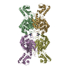
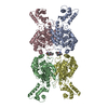
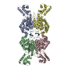
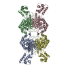
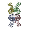
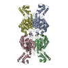
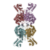
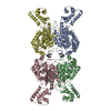
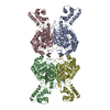
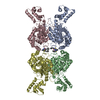
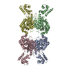
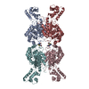
| Title | Crystal structure of glutaminase C in complex with inhibitor CB-839 | |||||||||
 Components Components | Glutaminase kidney isoform, mitochondrial | |||||||||
 Keywords Keywords | HYDROLASE/HYDROLASE INHIBITOR /  glutaminase / CB-839 / HYDROLASE-HYDROLASE INHIBITOR complex glutaminase / CB-839 / HYDROLASE-HYDROLASE INHIBITOR complex | |||||||||
| Function / homology |  Function and homology information Function and homology informationglutamine catabolic process / glutamate biosynthetic process / regulation of respiratory gaseous exchange by nervous system process / Glutamate and glutamine metabolism / intracellular glutamate homeostasis / Glutamate Neurotransmitter Release Cycle / glutaminase / glutaminase activity / suckling behavior / TP53 Regulates Metabolic Genes ...glutamine catabolic process / glutamate biosynthetic process / regulation of respiratory gaseous exchange by nervous system process / Glutamate and glutamine metabolism / intracellular glutamate homeostasis / Glutamate Neurotransmitter Release Cycle / glutaminase / glutaminase activity / suckling behavior / TP53 Regulates Metabolic Genes / chemical synaptic transmission / protein homotetramerization / mitochondrial matrix / synapse / mitochondrion / cytosolSimilarity search - Function | |||||||||
| Biological species |  Homo sapiens (human) Homo sapiens (human) | |||||||||
| Method | X-RAY DIFFRACTION / SYNCHROTRON / MOLECULAR REPLACEMENT / Resolution: 2.4 Å | |||||||||
 Authors Authors | Huang, Q. / Cerione, R.A. | |||||||||
| Funding support |  United States, 2items United States, 2items
| |||||||||
 Citation Citation | Journal: To Be Published Title: Crystal structure of the clinically relevant glutaminase inhibitot CB-839 in complex with glutaminase C Authors: Huang, Q. / Katt, W.P. / McDermott, L.A. / Cerione, R.A. | |||||||||
| History |
|
- Structure visualization
| Structure viewer | Molecule: MolmilJmol/JSmol |
|---|
- Downloads & links
-Download
| PDBx/mmCIF format | 5hl1.cif.gz | 334.3 KB | Display | PDBx/mmCIF format |
|---|---|---|---|---|
| PDB format | pdb5hl1.ent.gz | 267.9 KB | Display | PDB format |
| PDBx/mmJSON format | 5hl1.json.gz | Tree view | PDBx/mmJSON format | |
| Others |  Other downloads Other downloads |
-Validation report
| Arichive directory | https://data.pdbj.org/pub/pdb/validation_reports/hl/5hl1ftp://data.pdbj.org/pub/pdb/validation_reports/hl/5hl1 | HTTPS FTP |
|---|
-Related structure data
| Related structure data | 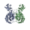 5d3oS S: Starting model for refinement |
|---|---|
| Similar structure data |


-Links
 PDBj
PDBj
- Assembly
| Deposited unit | 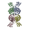
| ||||||||
|---|---|---|---|---|---|---|---|---|---|
| 1 |
| ||||||||
| Unit cell |
|
-Components
| #1: Protein | Mass: 59457.590 Da / Num. of mol.: 4 Source method: isolated from a genetically manipulated source Source: (gene. exp.) Homo sapiens (human) / Gene: GLS, GLS1, KIAA0838 / Plasmid: pQE80 / Production host:  Escherichia coli (E. coli) / Strain (production host): BL21(DE3) / References: UniProt: O94925, glutaminase Escherichia coli (E. coli) / Strain (production host): BL21(DE3) / References: UniProt: O94925, glutaminase#2: Chemical |   Mass: 571.574 Da / Num. of mol.: 2 / Source method: obtained synthetically / Formula: C26H24F3N7O3S Mass: 571.574 Da / Num. of mol.: 2 / Source method: obtained synthetically / Formula: C26H24F3N7O3S#3: Water | ChemComp-HOH / | Water Mass: 18.015 Da / Num. of mol.: 422 / Source method: isolated from a natural source / Formula: H2O Mass: 18.015 Da / Num. of mol.: 422 / Source method: isolated from a natural source / Formula: H2O |
|---|
-Experimental details
-Experiment
| Experiment | Method: X-RAY DIFFRACTION |
|---|
- Sample preparation
| Crystal | Density Matthews: 2.57 Å3/Da / Density % sol: 52.14 % |
|---|---|
| Crystal grow | Temperature: 293 K / Method: vapor diffusion, hanging drop / pH: 8 / Details: 10% PEG6000 / PH range: 8.0-8.5 |
-Data collection
| Diffraction | Mean temperature: 100 K |
|---|---|
| Diffraction source | Source: SYNCHROTRON / Site: CHESS / Beamline: A1 / Wavelength: 0.63 Å |
| Detector | Type: ADSC QUANTUM 210 / Detector: CCD / Date: May 28, 2015 |
| Radiation | Protocol: SINGLE WAVELENGTH / Monochromatic (M) / Laue (L): M / Scattering type: x-ray |
| Radiation wavelength | Wavelength: 0.63 Å / Relative weight: 1 |
| Reflection | Resolution: 2.4→50 Å / Num. obs: 96364 / % possible obs: 99.9 % / Redundancy: 7.3 % / Net I/σ(I): 15.6 |
- Processing
| Software |
| |||||||||||||||||||||||||||||||||||||||||||||||||||||||||||||||||||||||||||||||||||||||||||||||||||||||||
|---|---|---|---|---|---|---|---|---|---|---|---|---|---|---|---|---|---|---|---|---|---|---|---|---|---|---|---|---|---|---|---|---|---|---|---|---|---|---|---|---|---|---|---|---|---|---|---|---|---|---|---|---|---|---|---|---|---|---|---|---|---|---|---|---|---|---|---|---|---|---|---|---|---|---|---|---|---|---|---|---|---|---|---|---|---|---|---|---|---|---|---|---|---|---|---|---|---|---|---|---|---|---|---|---|---|---|
| Refinement | Method to determine structure: MOLECULAR REPLACEMENT Starting model: 5D3O Resolution: 2.4→47.479 Å / SU ML: 0.28 / Cross valid method: FREE R-VALUE / σ(F): 1.34 / Phase error: 32.05 / Stereochemistry target values: ML
| |||||||||||||||||||||||||||||||||||||||||||||||||||||||||||||||||||||||||||||||||||||||||||||||||||||||||
| Solvent computation | Shrinkage radii: 0.9 Å / VDW probe radii: 1.11 Å / Solvent model: FLAT BULK SOLVENT MODEL | |||||||||||||||||||||||||||||||||||||||||||||||||||||||||||||||||||||||||||||||||||||||||||||||||||||||||
| Refinement step | Cycle: LAST / Resolution: 2.4→47.479 Å
| |||||||||||||||||||||||||||||||||||||||||||||||||||||||||||||||||||||||||||||||||||||||||||||||||||||||||
| Refine LS restraints |
| |||||||||||||||||||||||||||||||||||||||||||||||||||||||||||||||||||||||||||||||||||||||||||||||||||||||||
| LS refinement shell |
|
