 Movie
Movie Controller

[English] 日本語
 Yorodumi
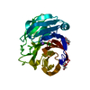
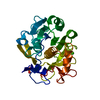
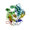
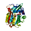
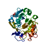
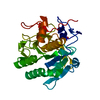
- PDB-5hij: Crystal structure of glycine sarcosine N-methyltransferase from M... -
+ Open data

- Basic information
| Entry | Database: PDB / ID: 5hij | ||||||||||||
|---|---|---|---|---|---|---|---|---|---|---|---|---|---|
| Title | Crystal structure of glycine sarcosine N-methyltransferase from Methanohalophilus portucalensis in complex with betaine | ||||||||||||
 Components Components | Glycine sarcosine N-methyltransferase | ||||||||||||
 Keywords Keywords |  TRANSFERASE / S-adenosylmethionine-dependent methyltransferases (SAM or AdoMet-MTase) / class I / monomethylation of glycine and sarcosine / rate-limiting enzyme in betaine biosynthesis / betaine-mediated feedback inhibition TRANSFERASE / S-adenosylmethionine-dependent methyltransferases (SAM or AdoMet-MTase) / class I / monomethylation of glycine and sarcosine / rate-limiting enzyme in betaine biosynthesis / betaine-mediated feedback inhibition | ||||||||||||
| Function / homology |  Function and homology information Function and homology informationcellular nitrogen compound metabolic process / glycine N-methyltransferase activity / organic substance biosynthetic process / cellular biosynthetic process / methylationSimilarity search - Function | ||||||||||||
| Biological species |  Methanohalophilus portucalensis FDF-1 (archaea) Methanohalophilus portucalensis FDF-1 (archaea) | ||||||||||||
| Method | X-RAY DIFFRACTION / SYNCHROTRON / MOLECULAR REPLACEMENT / Resolution: 1.93 Å | ||||||||||||
 Authors Authors | Lee, Y.R. / Lin, T.S. / Lai, S.J. / Liu, M.S. / Lai, M.C. / Chan, N.L. | ||||||||||||
| Funding support |  Taiwan, 3items Taiwan, 3items
| ||||||||||||
 Citation Citation | Journal: Sci Rep / Year: 2016 Title: Structural Analysis of Glycine Sarcosine N-methyltransferase from Methanohalophilus portucalensis Reveals Mechanistic Insights into the Regulation of Methyltransferase Activity Authors: Lee, Y.R. / Lin, T.S. / Lai, S.J. / Liu, M.S. / Lai, M.C. / Chan, N.L. | ||||||||||||
| History |
|
- Structure visualization
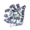
| Structure viewer | Molecule: MolmilJmol/JSmol |
|---|
- Downloads & links
-Download
| PDBx/mmCIF format | 5hij.cif.gz | 118.9 KB | Display | PDBx/mmCIF format |
|---|---|---|---|---|
| PDB format | pdb5hij.ent.gz | 89.4 KB | Display | PDB format |
| PDBx/mmJSON format | 5hij.json.gz | Tree view | PDBx/mmJSON format | |
| Others |  Other downloads Other downloads |
-Validation report
| Arichive directory | https://data.pdbj.org/pub/pdb/validation_reports/hi/5hijftp://data.pdbj.org/pub/pdb/validation_reports/hi/5hij | HTTPS FTP |
|---|
-Related structure data
| Related structure data |  5gwxC 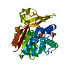 5h02C  5hiiC  5hikC  5hilSC  5himC C: citing same article ( S: Starting model for refinement |
|---|---|
| Similar structure data |


-Links
 PDBj
PDBj- Assembly
| Deposited unit | 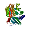
| ||||||||||||||||||
|---|---|---|---|---|---|---|---|---|---|---|---|---|---|---|---|---|---|---|---|
| 1 |
| ||||||||||||||||||
| Unit cell |
| ||||||||||||||||||
| Components on special symmetry positions |
|
-Components
| #1: Protein | Mass: 32800.289 Da / Num. of mol.: 1 Source method: isolated from a genetically manipulated source Source: (gene. exp.) Methanohalophilus portucalensis FDF-1 (archaea)Strain: FDF-1 / Gene: gsmt / Production host:  Escherichia coli BL21(DE3) (bacteria) / Strain (production host): BL21(DE3) / References: UniProt: F6KV61, EC: 2.1.1.156 Escherichia coli BL21(DE3) (bacteria) / Strain (production host): BL21(DE3) / References: UniProt: F6KV61, EC: 2.1.1.156 |
|---|---|
| #2: Chemical | ChemComp-BET / Trimethylglycine  Mass: 118.154 Da / Num. of mol.: 1 / Source method: obtained synthetically / Formula: C5H12NO2 Mass: 118.154 Da / Num. of mol.: 1 / Source method: obtained synthetically / Formula: C5H12NO2 |
| #3: Water | ChemComp-HOH / Water Mass: 18.015 Da / Num. of mol.: 190 / Source method: isolated from a natural source / Formula: H2O Mass: 18.015 Da / Num. of mol.: 190 / Source method: isolated from a natural source / Formula: H2O |
-Experimental details
-Experiment
| Experiment | Method: X-RAY DIFFRACTION / Number of used crystals: 1 |
|---|
- Sample preparation
| Crystal | Density Matthews: 3.15 Å3/Da / Density % sol: 60.94 % |
|---|---|
| Crystal grow | Temperature: 277.15 K / Method: vapor diffusion, hanging drop Details: Protein solution: GSMT (6.6 mg/ml) in 100 mM TES pH 7.3, 2 M KCl, 1 mM EDTA and 1 mM 2-Mercaptoethanol. Crystallization reagent: 0.2 M Potassium sodium tartrate tetrahydrate, 20% w/v ...Details: Protein solution: GSMT (6.6 mg/ml) in 100 mM TES pH 7.3, 2 M KCl, 1 mM EDTA and 1 mM 2-Mercaptoethanol. Crystallization reagent: 0.2 M Potassium sodium tartrate tetrahydrate, 20% w/v Polyethylene glycol 3,350. Betaine was soaked into crystals before data collection. |
-Data collection
| Diffraction | Mean temperature: 100 K |
|---|---|
| Diffraction source | Source: SYNCHROTRON / Site: NSRRC / Beamline: BL15A1 / Wavelength: 1 Å |
| Detector | Type: RAYONIX MX300HE / Detector: CCD / Date: Jun 27, 2015 |
| Radiation | Monochromator: LN2-Cooled, Fixed-Exit Double Crystal Monochromator Protocol: SINGLE WAVELENGTH / Monochromatic (M) / Laue (L): M / Scattering type: x-ray |
| Radiation wavelength | Wavelength: 1 Å / Relative weight: 1 |
| Reflection | Resolution: 1.93→20 Å / Num. obs: 31210 / % possible obs: 99.8 % / Redundancy: 4.3 % / Rsym value: 0.054 / Net I/σ(I): 21.7 |
| Reflection shell | Resolution: 1.93→2 Å / Redundancy: 4.3 % / Mean I/σ(I) obs: 3.3 / % possible all: 100 |
- Processing
| Software |
| |||||||||||||||||||||||||||||||||||||||||||||||||||||||||||||||||||||||||||||||||||||||||||||||||||||||||||||||||||||||||||||
|---|---|---|---|---|---|---|---|---|---|---|---|---|---|---|---|---|---|---|---|---|---|---|---|---|---|---|---|---|---|---|---|---|---|---|---|---|---|---|---|---|---|---|---|---|---|---|---|---|---|---|---|---|---|---|---|---|---|---|---|---|---|---|---|---|---|---|---|---|---|---|---|---|---|---|---|---|---|---|---|---|---|---|---|---|---|---|---|---|---|---|---|---|---|---|---|---|---|---|---|---|---|---|---|---|---|---|---|---|---|---|---|---|---|---|---|---|---|---|---|---|---|---|---|---|---|---|
| Refinement | Method to determine structure: MOLECULAR REPLACEMENT Starting model: PDB ENTRY 5HIL Resolution: 1.93→19.724 Å / SU ML: 0.2 / Cross valid method: FREE R-VALUE / σ(F): 1.35 / Phase error: 19.29 / Stereochemistry target values: ML
| |||||||||||||||||||||||||||||||||||||||||||||||||||||||||||||||||||||||||||||||||||||||||||||||||||||||||||||||||||||||||||||
| Solvent computation | Shrinkage radii: 0.9 Å / VDW probe radii: 1.11 Å / Solvent model: FLAT BULK SOLVENT MODEL | |||||||||||||||||||||||||||||||||||||||||||||||||||||||||||||||||||||||||||||||||||||||||||||||||||||||||||||||||||||||||||||
| Refinement step | Cycle: LAST / Resolution: 1.93→19.724 Å
| |||||||||||||||||||||||||||||||||||||||||||||||||||||||||||||||||||||||||||||||||||||||||||||||||||||||||||||||||||||||||||||
| Refine LS restraints |
| |||||||||||||||||||||||||||||||||||||||||||||||||||||||||||||||||||||||||||||||||||||||||||||||||||||||||||||||||||||||||||||
| LS refinement shell |
| |||||||||||||||||||||||||||||||||||||||||||||||||||||||||||||||||||||||||||||||||||||||||||||||||||||||||||||||||||||||||||||
| Refinement TLS params. | Method: refined / Refine-ID: X-RAY DIFFRACTION
| |||||||||||||||||||||||||||||||||||||||||||||||||||||||||||||||||||||||||||||||||||||||||||||||||||||||||||||||||||||||||||||
| Refinement TLS group |
|
