 Movie
Movie Controller


+ Open data

- Basic information
| Entry | Database: PDB / ID: 5h9q | ||||||||||||
|---|---|---|---|---|---|---|---|---|---|---|---|---|---|
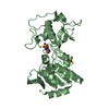
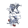
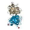
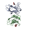
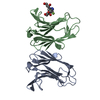
| Title | Crystal Structure of Human Galectin-7 in Complex with TD139 | ||||||||||||
 Components Components | Galectin-7 | ||||||||||||
 Keywords Keywords | SUGAR BINDING PROTEIN / galectin / thio-digalactoside (TD139) / pi-arginine interaction / fluorine bonding | ||||||||||||
| Function / homology |  Function and homology information Function and homology informationheterophilic cell-cell adhesion via plasma membrane cell adhesion molecules / carbohydrate binding / apoptotic process / extracellular space / extracellular exosome / nucleus / cytoplasmSimilarity search - Function | ||||||||||||
| Biological species |  Homo sapiens (human) Homo sapiens (human) | ||||||||||||
| Method | X-RAY DIFFRACTION / SYNCHROTRON / MOLECULAR REPLACEMENT / Resolution: 1.931 Å | ||||||||||||
 Authors Authors | Hsieh, T.J. / Lin, H.Y. / Lin, C.H. | ||||||||||||
| Funding support |  Taiwan, 3items Taiwan, 3items
| ||||||||||||
 Citation Citation | Journal: Sci Rep / Year: 2016 Title: Dual thio-digalactoside-binding modes of human galectins as the structural basis for the design of potent and selective inhibitors Authors: Hsieh, T.J. / Lin, H.Y. / Tu, Z. / Lin, T.C. / Wu, S.C. / Tseng, Y.Y. / Liu, F.T. / Danny Hsu, S.T. / Lin, C.H. | ||||||||||||
| History |
|
- Structure visualization
| Structure viewer | Molecule: MolmilJmol/JSmol |
|---|
- Downloads & links
-Download
| PDBx/mmCIF format | 5h9q.cif.gz | 75.7 KB | Display | PDBx/mmCIF format |
|---|---|---|---|---|
| PDB format | pdb5h9q.ent.gz | 53.8 KB | Display | PDB format |
| PDBx/mmJSON format | 5h9q.json.gz | Tree view | PDBx/mmJSON format | |
| Others |  Other downloads Other downloads |
-Validation report
| Arichive directory | https://data.pdbj.org/pub/pdb/validation_reports/h9/5h9qftp://data.pdbj.org/pub/pdb/validation_reports/h9/5h9q | HTTPS FTP |
|---|
-Related structure data
| Related structure data |  4y24C 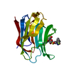 5h9pC  5h9rC  5h9sC 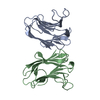 1bkzS C: citing same article ( S: Starting model for refinement |
|---|---|
| Similar structure data |


-Links
 PDBj
PDBj- Assembly
| Deposited unit | 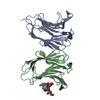
| ||||||||
|---|---|---|---|---|---|---|---|---|---|
| 1 |
| ||||||||
| Unit cell |
|
-Components
| #1: Protein | / Gal-7 / HKL-14 / PI7 / p53-induced gene 1 protein Mass: 17137.221 Da / Num. of mol.: 2 Source method: isolated from a genetically manipulated source Source: (gene. exp.) Homo sapiens (human) / Gene: LGALS7, PIG1, LGALS7B / Plasmid: pET28aProduction host:  Escherichia coli 'BL21-Gold(DE3)pLysS AG' (bacteria) Escherichia coli 'BL21-Gold(DE3)pLysS AG' (bacteria)Strain (production host): BL21-Gold(DE3)pLysS AG / References: UniProt: P47929 #2: Chemical | ChemComp-TD2 / |   Mass: 648.635 Da / Num. of mol.: 1 / Source method: obtained synthetically / Formula: C28H30F2N6O8S Mass: 648.635 Da / Num. of mol.: 1 / Source method: obtained synthetically / Formula: C28H30F2N6O8S#3: Water | ChemComp-HOH / | Water Mass: 18.015 Da / Num. of mol.: 256 / Source method: isolated from a natural source / Formula: H2O Mass: 18.015 Da / Num. of mol.: 256 / Source method: isolated from a natural source / Formula: H2O |
|---|
-Experimental details
-Experiment
| Experiment | Method: X-RAY DIFFRACTION |
|---|
- Sample preparation
| Crystal | Density Matthews: 1.79 Å3/Da / Density % sol: 31.23 % |
|---|---|
| Crystal grow | Temperature: 298 K / Method: vapor diffusion, hanging drop / pH: 8 / Details: 0.22 M Mg Acetate, 21% (w/v) PEG3350 / PH range: 7.5-8.5 |
-Data collection
| Diffraction | Mean temperature: 100 K |
|---|---|
| Diffraction source | Source: SYNCHROTRON / Site: NSRRC / Beamline: BL15A1 / Wavelength: 1 Å |
| Detector | Type: ADSC QUANTUM 315r / Detector: CCD / Date: May 24, 2013 |
| Radiation | Protocol: SINGLE WAVELENGTH / Monochromatic (M) / Laue (L): M / Scattering type: x-ray |
| Radiation wavelength | Wavelength: 1 Å / Relative weight: 1 |
| Reflection | Resolution: 1.93→30 Å / Num. obs: 19082 / % possible obs: 99.6 % / Redundancy: 6 % / Rmerge(I) obs: 0.036 / Net I/σ(I): 44.9 |
| Reflection shell | Resolution: 1.93→2 Å / Redundancy: 6 % / Rmerge(I) obs: 0.152 / Mean I/σ(I) obs: 11.86 / % possible all: 96.5 |
- Processing
| Software |
| |||||||||||||||||||||||||||||||||||||||||||||||||||||||||||||||||||||||||||||||||||||||||||||||||||||||||
|---|---|---|---|---|---|---|---|---|---|---|---|---|---|---|---|---|---|---|---|---|---|---|---|---|---|---|---|---|---|---|---|---|---|---|---|---|---|---|---|---|---|---|---|---|---|---|---|---|---|---|---|---|---|---|---|---|---|---|---|---|---|---|---|---|---|---|---|---|---|---|---|---|---|---|---|---|---|---|---|---|---|---|---|---|---|---|---|---|---|---|---|---|---|---|---|---|---|---|---|---|---|---|---|---|---|---|
| Refinement | Method to determine structure: MOLECULAR REPLACEMENT Starting model: 1BKZ Resolution: 1.931→27.651 Å / SU ML: 0.18 / Cross valid method: FREE R-VALUE / σ(F): 0 / Phase error: 22.71 / Stereochemistry target values: ML
| |||||||||||||||||||||||||||||||||||||||||||||||||||||||||||||||||||||||||||||||||||||||||||||||||||||||||
| Solvent computation | Shrinkage radii: 0.9 Å / VDW probe radii: 1.11 Å / Solvent model: FLAT BULK SOLVENT MODEL | |||||||||||||||||||||||||||||||||||||||||||||||||||||||||||||||||||||||||||||||||||||||||||||||||||||||||
| Refinement step | Cycle: LAST / Resolution: 1.931→27.651 Å
| |||||||||||||||||||||||||||||||||||||||||||||||||||||||||||||||||||||||||||||||||||||||||||||||||||||||||
| Refine LS restraints |
| |||||||||||||||||||||||||||||||||||||||||||||||||||||||||||||||||||||||||||||||||||||||||||||||||||||||||
| LS refinement shell |
|
