 Movie
Movie Controller


+ Open data

- Basic information
| Entry | Database: PDB / ID: 5h9p | |||||||||
|---|---|---|---|---|---|---|---|---|---|---|
| Title | Crystal Structure of Human Galectin-3 CRD in Complex with TD139 | |||||||||
 Components Components | Galectin-3 | |||||||||
 Keywords Keywords | SUGAR BINDING PROTEIN / galectin / thio-digalactoside (TDG) / pi-arginine interaction | |||||||||
| Function / homology |  Function and homology information Function and homology informationnegative regulation of protein tyrosine phosphatase activity / negative regulation of immunological synapse formation / RUNX2 regulates genes involved in differentiation of myeloid cells / negative regulation of T cell activation via T cell receptor contact with antigen bound to MHC molecule on antigen presenting cell / regulation of T cell apoptotic process / mononuclear cell migration / IgE binding / positive regulation of mononuclear cell migration / negative regulation of endocytosis / eosinophil chemotaxis ...negative regulation of protein tyrosine phosphatase activity / negative regulation of immunological synapse formation / RUNX2 regulates genes involved in differentiation of myeloid cells / negative regulation of T cell activation via T cell receptor contact with antigen bound to MHC molecule on antigen presenting cell / regulation of T cell apoptotic process / mononuclear cell migration / IgE binding / positive regulation of mononuclear cell migration / negative regulation of endocytosis / eosinophil chemotaxis / regulation of extrinsic apoptotic signaling pathway via death domain receptors / RUNX1 regulates transcription of genes involved in differentiation of myeloid cells / protein phosphatase inhibitor activity / negative regulation of T cell receptor signaling pathway / positive chemotaxis / macrophage chemotaxis / regulation of T cell proliferation / positive regulation of calcium ion import / chemoattractant activity / monocyte chemotaxis / Advanced glycosylation endproduct receptor signaling / ficolin-1-rich granule membrane / immunological synapse / laminin binding / epithelial cell differentiation / molecular condensate scaffold activity / neutrophil chemotaxis / RNA splicing / secretory granule membrane / positive regulation of protein-containing complex assembly / negative regulation of extrinsic apoptotic signaling pathway / positive regulation of protein localization to plasma membrane / spliceosomal complex / mRNA processing / carbohydrate binding / collagen-containing extracellular matrix / protein phosphatase binding / mitochondrial inner membrane / innate immune response / Neutrophil degranulation / cell surface / extracellular space / RNA binding / extracellular exosome / extracellular region / nucleoplasm / membrane / nucleus / plasma membrane / cytosol / cytoplasmSimilarity search - Function | |||||||||
| Biological species |  Homo sapiens (human) Homo sapiens (human) | |||||||||
| Method | X-RAY DIFFRACTION / SYNCHROTRON / MOLECULAR REPLACEMENT / Resolution: 2.04 Å | |||||||||
 Authors Authors | Hsieh, T.J. / Lin, H.Y. / Lin, C.H. | |||||||||
| Funding support |  Taiwan, 2items Taiwan, 2items
| |||||||||
 Citation Citation | Journal: Sci Rep / Year: 2016 Title: Dual thio-digalactoside-binding modes of human galectins as the structural basis for the design of potent and selective inhibitors Authors: Hsieh, T.J. / Lin, H.Y. / Tu, Z. / Lin, T.C. / Wu, S.C. / Tseng, Y.Y. / Liu, F.T. / Danny Hsu, S.T. / Lin, C.H. | |||||||||
| History |
|
- Structure visualization
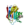
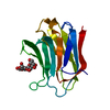
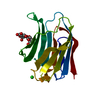
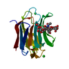
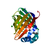
| Structure viewer | Molecule: MolmilJmol/JSmol |
|---|
- Downloads & links
-Download
| PDBx/mmCIF format | 5h9p.cif.gz | 47.8 KB | Display | PDBx/mmCIF format |
|---|---|---|---|---|
| PDB format | pdb5h9p.ent.gz | 30.2 KB | Display | PDB format |
| PDBx/mmJSON format | 5h9p.json.gz | Tree view | PDBx/mmJSON format | |
| Others |  Other downloads Other downloads |
-Validation report
| Arichive directory | https://data.pdbj.org/pub/pdb/validation_reports/h9/5h9pftp://data.pdbj.org/pub/pdb/validation_reports/h9/5h9p | HTTPS FTP |
|---|
-Related structure data
| Related structure data |  4y24C 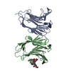 5h9qC  5h9rC 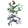 5h9sC  2nmnS C: citing same article ( S: Starting model for refinement |
|---|---|
| Similar structure data |


-Links
 PDBj
PDBj


- Assembly
| Deposited unit | 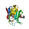
| ||||||||
|---|---|---|---|---|---|---|---|---|---|
| 1 |
| ||||||||
| Unit cell |
|
-Components
| #1: Protein | / Gal-3 / 35 kDa lectin / Carbohydrate-binding protein 35 / CBP 35 / Galactose-specific lectin 3 / ...Gal-3 / 35 kDa lectin / Carbohydrate-binding protein 35 / CBP 35 / Galactose-specific lectin 3 / Galactoside-binding protein / GALBP / IgE-binding protein / L-31 / Laminin-binding protein / Lectin L-29 / Mac-2 antigen Mass: 17872.418 Da / Num. of mol.: 1 Fragment: carbohydrate-recognition domain, UNP residues 113-250 Source method: isolated from a genetically manipulated source Source: (gene. exp.) Homo sapiens (human) / Gene: LGALS3, MAC2Production host:  Escherichia coli 'BL21-Gold(DE3)pLysS AG' (bacteria) Escherichia coli 'BL21-Gold(DE3)pLysS AG' (bacteria)Strain (production host): BL21-Gold(DE3)pLysS AG / References: UniProt: P17931 |
|---|---|
| #2: Chemical | ChemComp-TD2 /   Mass: 648.635 Da / Num. of mol.: 1 / Source method: obtained synthetically / Formula: C28H30F2N6O8S Mass: 648.635 Da / Num. of mol.: 1 / Source method: obtained synthetically / Formula: C28H30F2N6O8S |
| #3: Water | ChemComp-HOH / Water Mass: 18.015 Da / Num. of mol.: 84 / Source method: isolated from a natural source / Formula: H2O Mass: 18.015 Da / Num. of mol.: 84 / Source method: isolated from a natural source / Formula: H2O |
-Experimental details
-Experiment
| Experiment | Method: X-RAY DIFFRACTION / Number of used crystals: 1 |
|---|
- Sample preparation
| Crystal | Density Matthews: 1.89 Å3/Da / Density % sol: 34.96 % |
|---|---|
| Crystal grow | Temperature: 298 K / Method: vapor diffusion, hanging drop / pH: 8 / Details: 0.1 M Tris pH 8.0, 0.2 M LiSO4, 30% (w/v) PEG 4000 / PH range: 7.5-8.5 |
-Data collection
| Diffraction | Mean temperature: 100 K | |||||||||||||||||||||||||||||||||||||||||||||||||||||||||||||||||||||||||||||||||||||||||||||||||||
|---|---|---|---|---|---|---|---|---|---|---|---|---|---|---|---|---|---|---|---|---|---|---|---|---|---|---|---|---|---|---|---|---|---|---|---|---|---|---|---|---|---|---|---|---|---|---|---|---|---|---|---|---|---|---|---|---|---|---|---|---|---|---|---|---|---|---|---|---|---|---|---|---|---|---|---|---|---|---|---|---|---|---|---|---|---|---|---|---|---|---|---|---|---|---|---|---|---|---|---|---|
| Diffraction source | Source: SYNCHROTRON / Site: NSRRC / Beamline: BL13B1 / Wavelength: 1 Å | |||||||||||||||||||||||||||||||||||||||||||||||||||||||||||||||||||||||||||||||||||||||||||||||||||
| Detector | Type: ADSC QUANTUM 315r / Detector: CCD / Date: Nov 17, 2013 | |||||||||||||||||||||||||||||||||||||||||||||||||||||||||||||||||||||||||||||||||||||||||||||||||||
| Radiation | Protocol: SINGLE WAVELENGTH / Monochromatic (M) / Laue (L): M / Scattering type: x-ray | |||||||||||||||||||||||||||||||||||||||||||||||||||||||||||||||||||||||||||||||||||||||||||||||||||
| Radiation wavelength | Wavelength: 1 Å / Relative weight: 1 | |||||||||||||||||||||||||||||||||||||||||||||||||||||||||||||||||||||||||||||||||||||||||||||||||||
| Reflection | Resolution: 2.04→30 Å / Num. obs: 9047 / % possible obs: 99 % / Redundancy: 10.5 % / Rmerge(I) obs: 0.071 / Rpim(I) all: 0.022 / Rrim(I) all: 0.074 / Χ2: 1.021 / Net I/av σ(I): 30.541 / Net I/σ(I): 15.2 / Num. measured all: 95013 | |||||||||||||||||||||||||||||||||||||||||||||||||||||||||||||||||||||||||||||||||||||||||||||||||||
| Reflection shell | Diffraction-ID: 1 / Rejects: 0
|
- Processing
| Software |
| |||||||||||||||||||||||||||||||||||||||||||||||||
|---|---|---|---|---|---|---|---|---|---|---|---|---|---|---|---|---|---|---|---|---|---|---|---|---|---|---|---|---|---|---|---|---|---|---|---|---|---|---|---|---|---|---|---|---|---|---|---|---|---|---|
| Refinement | Method to determine structure: MOLECULAR REPLACEMENT Starting model: 2NMN Resolution: 2.04→26.067 Å / SU ML: 0.2 / Cross valid method: FREE R-VALUE / σ(F): 0 / Phase error: 21.4 / Stereochemistry target values: ML
| |||||||||||||||||||||||||||||||||||||||||||||||||
| Solvent computation | Shrinkage radii: 0.9 Å / VDW probe radii: 1.11 Å / Solvent model: FLAT BULK SOLVENT MODEL | |||||||||||||||||||||||||||||||||||||||||||||||||
| Displacement parameters | Biso max: 78.19 Å2 / Biso mean: 27.7415 Å2 / Biso min: 11.81 Å2 | |||||||||||||||||||||||||||||||||||||||||||||||||
| Refinement step | Cycle: final / Resolution: 2.04→26.067 Å
| |||||||||||||||||||||||||||||||||||||||||||||||||
| Refine LS restraints |
| |||||||||||||||||||||||||||||||||||||||||||||||||
| LS refinement shell | Refine-ID: X-RAY DIFFRACTION / Total num. of bins used: 6
|
